Pictures of Basal Cell Carcinoma (BCC) – Warning Signs

Helping You Recognise BCC
Pictures of Basal Cell Carcinoma
If you have noticed an unusual mark, bump, or sore on your skin and are searching for pictures to compare it against, you are not alone. Many people turn to images when they first notice a skin change, and it is a perfectly natural first step.
However, it is important to remember that no image can replace a clinical assessment — the only way to know for certain whether a lesion is a BCC is to have it examined by a specialist.
This article provides an overview of what basal cell carcinoma looks like, the different subtypes and where they commonly appear, and guidance on what to do if you spot something on your skin that concerns you.
What Does Basal Cell Carcinoma Look Like?
Basal cell carcinoma does not always look the same. Its appearance varies depending on the subtype, its location on the body, and how long it has been present. This is one of the reasons BCCs can be easy to overlook in the early stages.
That said, there are several visual features that are common across most types of BCC:
- A pearly, shiny, or translucent surface — often described as looking like a small pearl or a smooth pimple that never quite goes away
- Pink, red, or skin-coloured in tone
- Visible small blood vessels (known as telangiectasia) on or around the surface
- A tendency to bleed easily, even from minor contact
- A scab or sore that heals partially and then returns
- In some cases, an area that resembles a scar without any history of injury to that area of skin


Request an e-Consultation
Speak with Miss Rakhee Nayar from the comfort of your own home and receive expert advice on your skin concern, treatment options, and next steps.
Understanding How BCC Differs
Types of Basal Cell Carcinoma
There are several subtypes of BCC, each with a slightly different appearance. Understanding these differences can help you recognise what to look out for on your own skin.

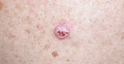
Nodular BCC
The most common subtype, nodular BCC typically appears as a round, raised bump with a pearly or translucent surface. It often has a rolled or defined border and may have small, visible blood vessels running across it. It is most commonly found on the face, particularly around the nose, cheeks, and forehead. In some cases, the centre of the lesion may break down to form a small ulcer.
Superficial BCC
Superficial BCC appears as a flat, red or pink scaly patch that can look similar to eczema or psoriasis. It tends to grow outwards rather than downwards, making it one of the easier subtypes to treat. It is most commonly found on the trunk, shoulders, and upper back.
Morphoeic or Infiltrative (Sclerosing) BCC
This is the most difficult subtype to detect and is often missed or misdiagnosed. Morphoeic BCC appears as a pale, flat, or slightly sunken patch that closely resembles a scar. Its borders are poorly defined, which also makes it one of the more challenging subtypes to treat surgically. It tends to infiltrate surrounding tissue more aggressively than other types.
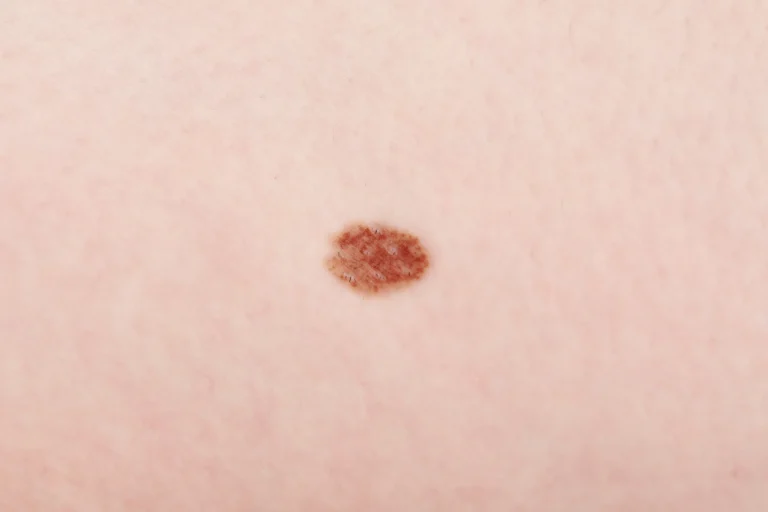
Pigmented BCC
Pigmented BCC shares many of the visual characteristics of nodular BCC but has areas of brown, blue, or black colouring within it. This can make it look similar to a melanoma, which is why specialist assessment and, if necessary, biopsy is essential for an accurate diagnosis.
Basosquamous BCC
This subtype shares features of both basal cell carcinoma and squamous cell carcinoma. It tends to behave more aggressively than other BCC subtypes and carries a slightly higher risk of spreading, making prompt treatment particularly important.
What Does BCC Look Like in Different Locations?
The appearance of a BCC can also vary depending on where it is located on the body. Here is what to look out for in the most common sites:

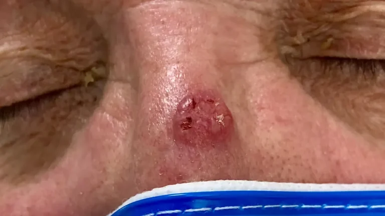
BCC on the face
The face is the most common location for BCC, particularly around the nose, eyes, cheeks, and forehead. Facial BCCs often present as a small pearly bump or a non-healing sore. Because of the proximity to critical structures such as the eyes and nose, facial BCCs require particularly careful treatment by an experienced specialist.

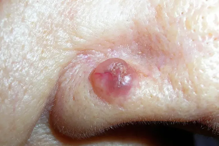
BCC on the Nose
The nose is one of the most frequently affected sites. BCCs here may appear as a shiny nodule, a scabbing sore, or a subtle flattened area that is easy to dismiss as dry skin. Treatment on the nose requires a precise technique to preserve both function and appearance.
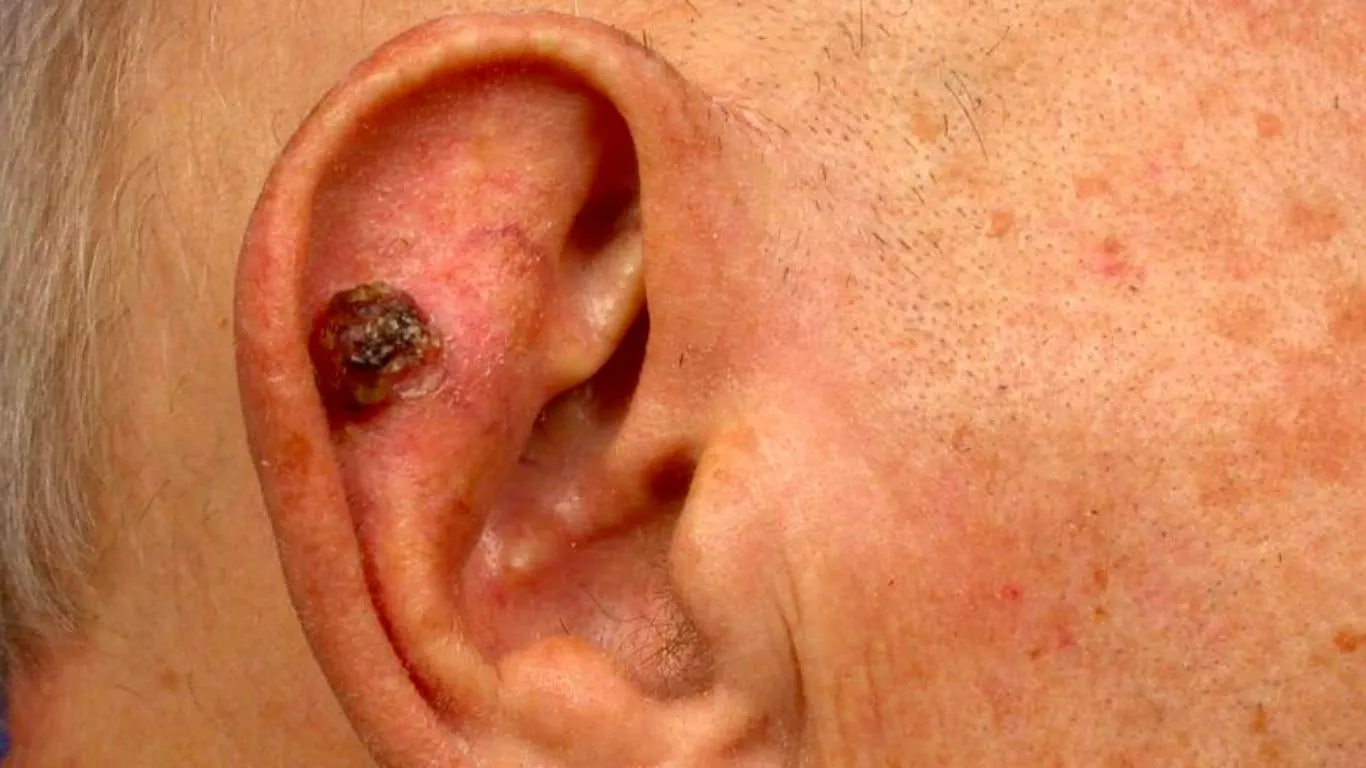
BCC on the ear
BCCs on the ear are often overlooked because the area is difficult to examine. They may present as a scaly or crusting patch, a persistent sore, or a small raised nodule on the outer ear or around the ear canal.
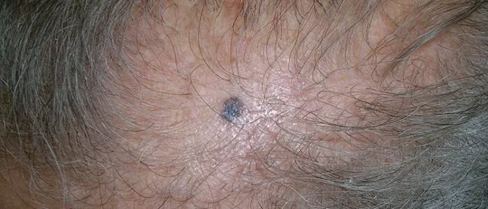
BCC on the scalp
Scalp BCCs can be hidden by hair and may only be noticed after repeated bleeding or a non-healing sore. Fair-skinned individuals with a history of significant sun exposure are at particular risk.
BCC on the back and shoulders
Superficial BCCs are more common on the trunk than other subtypes. They often appear as flat, scaly, red or pink patches and can be mistaken for areas of dry skin or mild eczema.
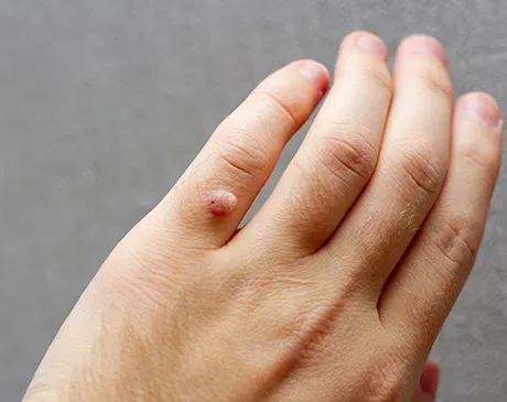
BCC on the hand
Whilst less common, BCC can develop on the hands and fingers. Treatment in this location requires particular care to preserve hand function. Miss Rakhee Nayar holds a European Hand Diploma (EBHS) and is one of very few surgeons in the UK with the specialist training to perform Mohs surgery on the hand, carefully limiting the surgical defect to maintain maximum function.
What Is the Difference Between Early and Advanced BCC?
The earlier a BCC is detected and treated, the simpler the procedure required and the better the outcome. Understanding the difference between early and advanced BCCs can help illustrate why prompt assessment matters.
Early stage BCC tends to be small, well-defined, and superficial. The borders are usually clear, tissue involvement is minimal, and treatment is straightforward. Many early BCCs can be treated with a relatively minor procedure under local anaesthetic.
Advanced BCC is larger, deeper, and may have poorly defined borders that make complete removal more technically demanding. Advanced BCCs are more likely to have invaded surrounding tissue and are more prone to recurrence following treatment. They may also require more extensive reconstruction after removal.
The key takeaway is simple: do not wait for a lesion to grow or change significantly before seeking advice. The sooner a BCC is assessed, the less invasive the treatment required.
Catch Suspicious Changes Early
How to Check Your Skin for BCC
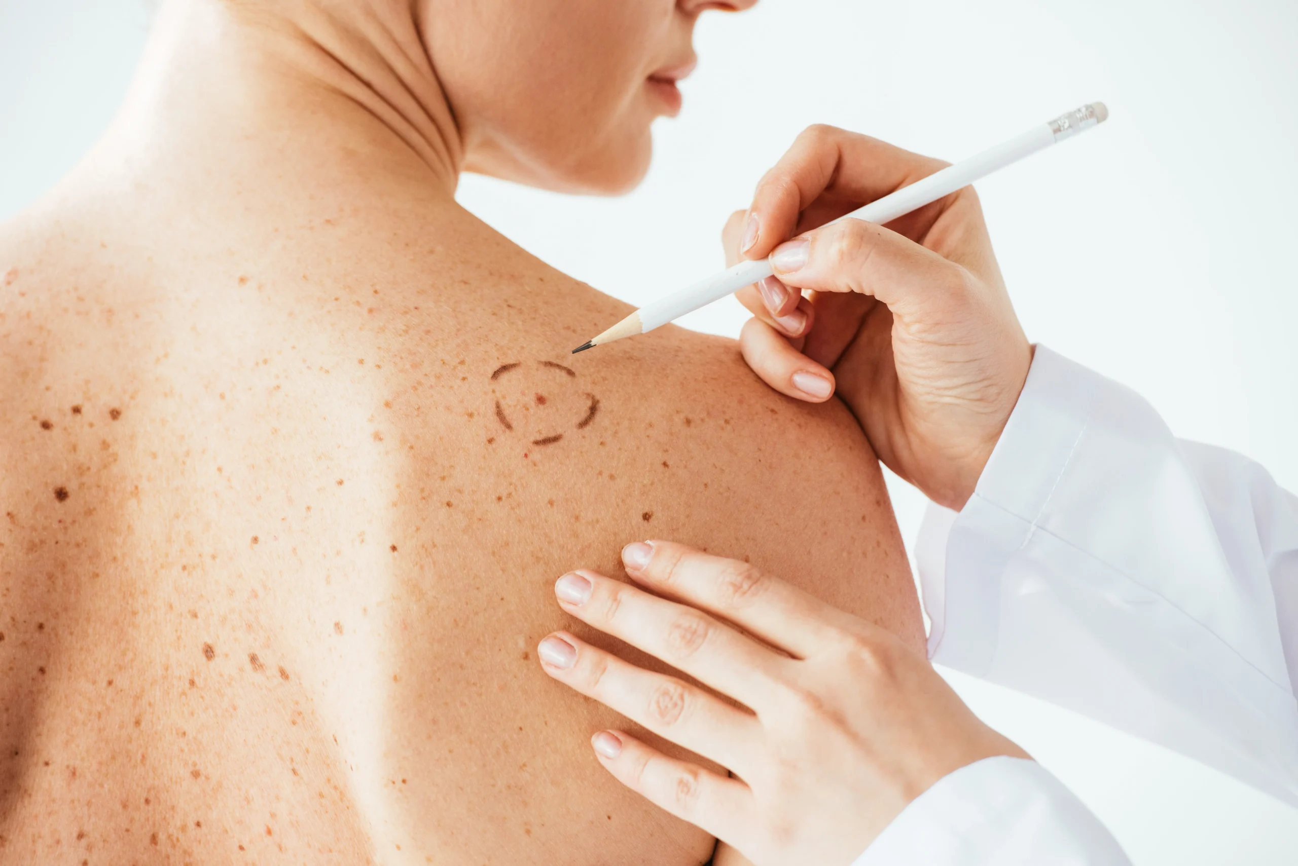
Regularly checking your skin is one of the most effective ways to catch a BCC early. When examining your skin, use the ABCDE checklist as a guide. If a lesion on your skin meets any of these criteria, do not wait to see if it resolves on its own. Book a specialist assessment as soon as possible.
Border: edges are poorly defined, rolled, or irregular
Diameter: any size warrants attention, but larger lesions should be assessed urgently
Evolution: any change in size, shape, colour, or a new symptom such as bleeding, itching, or crusting
For nodular BCC specifically, dermatologists also use the EFG rule as an additional guide:
E — Elevated: the lesion is raised above the surrounding skin
F — Firm: the lesion feels firm to the touch
G — Growing: the lesion has increased in size over time
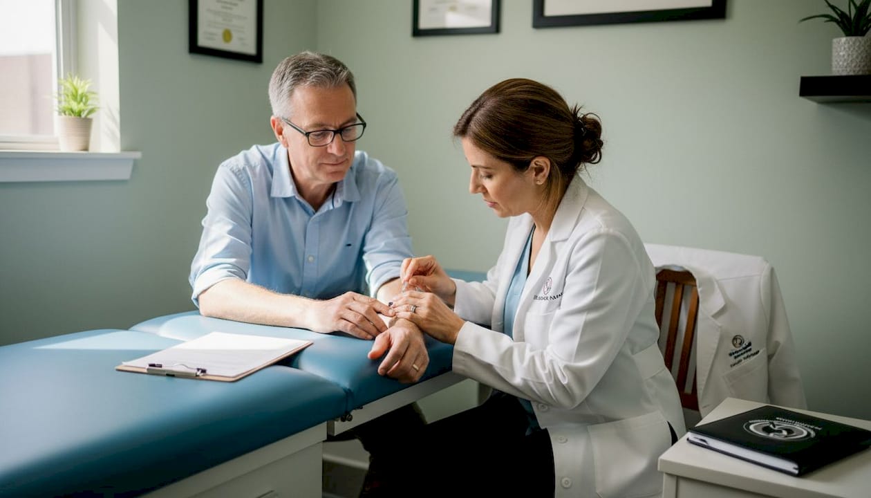
Concerned About a Lesion? See a Specialist
If any of the images or descriptions in this article look familiar, the most important step you can take is to have your lesion assessed by a specialist. A clinical examination, and if necessary a biopsy, is the only reliable way to confirm whether a lesion is a BCC and what treatment is needed.
Miss Rakhee Nayar offers prompt, expert assessment and surgical removal of BCCs across the North West, with e-Consultations available to patients throughout the UK.
As one of fewer than 10 women in the UK dual-trained in both plastic surgery and Mohs micrographic surgery, she brings over 20 years of experience and a 99.9% patient satisfaction rate to every consultation.

07740 306144
Frequently Asked Questions
Common Questions About BCC
Can you tell if a lesion is BCC just by looking at it?
Not with certainty. Whilst certain visual features are associated with BCC, the only reliable way to confirm a diagnosis is through a clinical assessment and, in most cases, a skin biopsy carried out by a specialist.
What is the most common appearance of BCC?
The most common type is nodular BCC, which typically appears as a small, round, pearly or translucent bump with a rolled border and visible blood vessels. It is most frequently found on the face.
Can BCC look like a pimple?
Yes. In its early stages, a nodular BCC can closely resemble a pimple or a small cyst. The key difference is that a BCC will not resolve on its own and may bleed, crust, or slowly grow over time.
Does BCC always bleed?
Not always, but bleeding is a common feature — particularly in nodular BCCs. Many patients first notice a BCC because it bleeds after minor contact, such as when washing or drying the face.
What is the difference between BCC and melanoma?
BCC and melanoma are both forms of skin cancer but they differ in origin, appearance, and behaviour. BCC develops in the basal cells and rarely spreads. Melanoma develops in the pigment-producing cells (melanocytes), tends to be darker in colour, and carries a significantly higher risk of spreading to other parts of the body. If you are unsure which type of lesion you have, always seek specialist assessment.
When should I see a doctor about a skin lesion?
You should seek a specialist opinion if a lesion is new, growing, changing in colour or shape, bleeding, crusting, or failing to heal. If you are in any doubt, it is always better to have a lesion reviewed sooner rather than later.