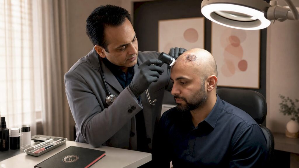
Skin cancer in the scalp is a keratinocyte malignancy arising most commonly as basal cell carcinoma (BCC) or squamous cell carcinoma (SCC) on the sun-exposed crown, temples, and hairline. The scalp receives cumulative ultraviolet (UV) radiation across a lifetime, yet hair cover delays detection, meaning lesions are often larger at presentation than equivalent tumours on the face. Rakhee Nayar – Mohs Surgeon and Skin Specialist sees this pattern regularly in clinical practice. Early recognition, prompt biopsy, and specialist surgical treatment are the three steps that determine outcome. This article explains each one clearly.
What are the symptoms of skin cancer in the scalp?
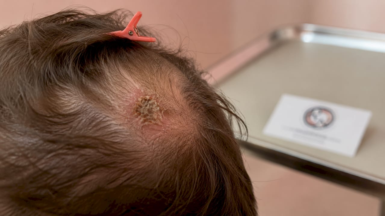
Persistent scalp lesions that do not heal, repeatedly crust, or ulcerate are the cardinal warning signs of BCC and SCC. The difficulty is that both cancers can mimic common benign conditions, including seborrhoeic keratoses, infected follicles, and eczema patches. That resemblance is precisely what delays diagnosis.
How BCC and SCC present differently
Basal cell carcinoma on the scalp typically appears as a shiny, pearlescent papule or nodule with a rolled edge and central depression. Over time it may ulcerate and bleed with minimal trauma. Squamous cell carcinoma presents differently: it tends to form a red, scaly plaque or a crusted papule that feels firm to the touch. SCC can grow faster than BCC and carries a higher metastasis risk if left untreated.
Key warning signs to watch for include:
- A sore or scab on the scalp that heals partially, then returns within weeks
- A shiny or translucent bump, often with visible small blood vessels
- A red, rough, or scaly patch that persists beyond four weeks
- A raised, firm nodule that bleeds easily when combed or brushed
- Any lesion that changes in size, colour, or texture over weeks to months
Pro Tip: Ask your hairdresser or barber to alert you to any unusual patches or lumps on your scalp at each appointment. They often notice changes before you do.
The most misleading presentation is the lesion that appears to heal. BCC in particular can crust over, giving the impression of recovery, only to re-open. Nonmelanoma scalp cancers are frequently dismissed for this reason, reducing the urgency for biopsy and delaying diagnosis by months or even years.
What are the risk factors for scalp skin cancer?
UV radiation is the primary cause of scalp skin cancer. The scalp is one of the most sun-exposed surfaces on the body, particularly in individuals with thinning hair, a shaved head, or a receding hairline. Chronic cumulative exposure, rather than a single severe sunburn, drives the majority of cases.
Personal risk factors that increase susceptibility include:
- Fair skin, light hair, and blue or green eyes, which reduce natural UV protection
- A history of sunburns, especially blistering burns before the age of 18
- Age over 50, as UV damage accumulates over decades
- A personal or family history of skin cancer
- Immunosuppression, including long-term steroid use or organ transplant medication
- Occupational or recreational outdoor exposure over many years
Solar keratosis (actinic keratosis) is a common precursor lesion on the scalp, particularly in bald men. It presents as a rough, scaly patch and represents dysplastic change driven by chronic sun damage. Left untreated, a proportion of solar keratoses progress to SCC.
Hair coverage creates a false sense of protection. Thick hair does reduce UV penetration, but a parting, a thinning crown, or a shaved head offers little barrier. Many patients are surprised to learn their scalp has received significant UV exposure across decades of outdoor activity without sun protection. Wearing a broad-brimmed hat and applying SPF 30 or higher sunscreen to exposed scalp skin are the most effective preventive measures available.
How is scalp skin cancer diagnosed?
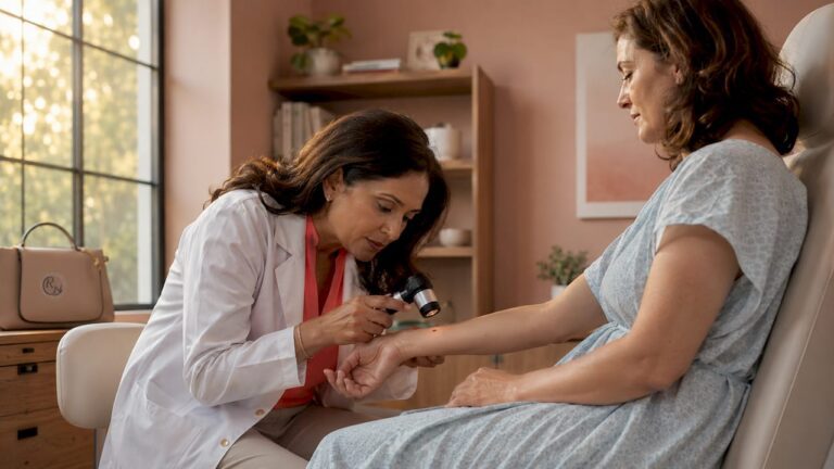
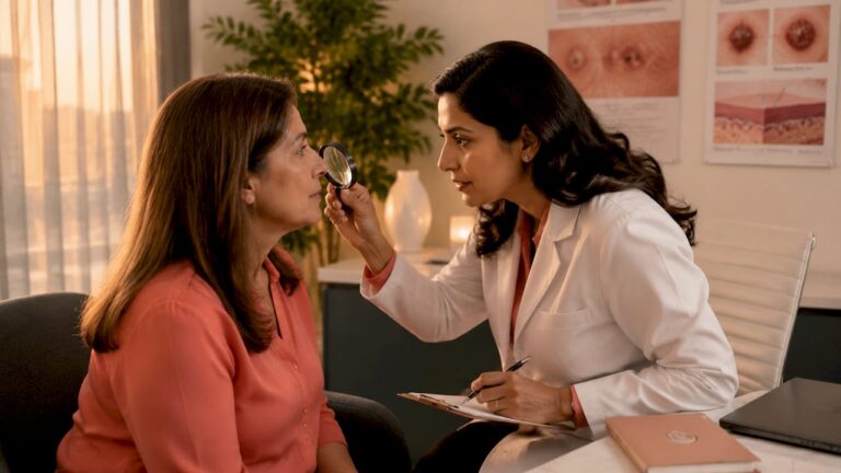
Visual inspection alone is insufficient to diagnose scalp skin cancer. Dermoscopy, a non-invasive technique using a handheld magnifying device with polarised light, improves diagnostic accuracy over unaided assessment and is particularly useful for recognising BCC and melanoma features before biopsy. However, dermoscopy does not replace tissue sampling.
Biopsy with histopathology is the gold standard for definitive diagnosis. The method chosen depends on lesion size, depth, and the clinical suspicion of subtype.
Common biopsy methods used on the scalp
- Shave biopsy: A superficial technique suited to raised, exophytic lesions. Quick and well-tolerated, but may not capture deeper tumour architecture.
- Punch biopsy: A circular blade removes a full-thickness core of skin, typically 3–6 mm in diameter. Preferred for flat or pigmented lesions where depth assessment matters.
- Incisional biopsy: A scalpel removes a representative portion of a larger lesion. Used when the full lesion cannot be excised at the diagnostic stage.
- Excisional biopsy: The entire lesion is removed with a margin of normal tissue. Appropriate for smaller lesions where complete removal and diagnosis can be achieved simultaneously.
Biopsy technique selection on the scalp is nuanced. Clinicians tailor the method according to depth, size, and the need for subtype diagnosis to prevent sampling error. Subtype matters because, for example, morphoeic BCC behaves more aggressively than nodular BCC and requires wider surgical margins.
| Biopsy Type | Best Used For | Limitation |
|---|---|---|
| Shave | Raised, exophytic lesions | May miss deep tumour extent |
| Punch | Flat or pigmented lesions | Small sample may not represent whole lesion |
| Incisional | Large lesions needing partial sampling | Does not provide definitive margin status |
| Excisional | Small lesions, complete removal | Not suitable for large or ill-defined tumours |
Pro Tip: If a GP or clinician suggests “watching and waiting” for a scalp lesion that has been present for more than six weeks without healing, ask specifically whether a biopsy referral is appropriate. Early tissue diagnosis prevents delays.
Once histopathology confirms the cancer type and subtype, treatment planning can begin. The report guides surgical margin requirements, the need for Mohs surgery, and whether adjunct therapies are indicated.
What treatment options are available for scalp skin cancer?
Treatment selection depends on tumour location, size, subtype, and patient priorities including functional and cosmetic outcomes. Surgery is the first-line treatment for the majority of scalp skin cancers.
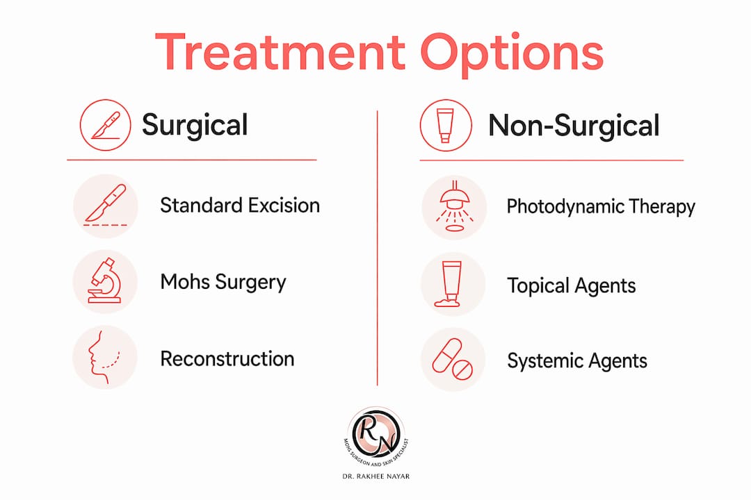
Surgical options
Surgical excision removes the tumour with a margin of normal tissue. Standard excision is appropriate for many primary, well-defined tumours. Mohs micrographic surgery is the preferred approach for complete tumour removal with tissue sparing, particularly where margins are difficult to define clinically or where the tumour is recurrent, large, or in a location where preserving healthy scalp tissue matters.
Mohs surgery examines tumour margins layer by layer in real time. Each layer is processed and mapped histologically before the next is removed. Surgery stops the moment clear margins are confirmed. This approach achieves the highest cure rates for BCC and SCC while removing the least amount of healthy tissue. For scalp tumours, where reconstruction can be complex, that tissue preservation is clinically significant.
Mohs surgery is favoured for scalp cancers specifically because it maximises margin control and minimises healthy tissue loss, balancing cure with cosmetic outcomes.
Non-surgical and adjunct treatments
| Treatment | Indication | Notes |
|---|---|---|
| Photodynamic therapy (PDT) | Superficial BCC, actinic keratosis | Not suitable for nodular or deep tumours |
| Topical 5-fluorouracil | Superficial BCC, actinic keratosis | Requires weeks of application; local reaction expected |
| Topical imiquimod | Superficial BCC | Immune-modulating agent; less reliable than surgery |
| Radiotherapy | Inoperable tumours or post-surgical adjuvant | Used when surgery is contraindicated or to reduce recurrence risk |
| Systemic therapy | Advanced or metastatic SCC or BCC | Hedgehog pathway inhibitors (vismodegib) for advanced BCC; immunotherapy for advanced SCC |
Photodynamic therapy and topical agents are typically reserved for superficial or early scalp cancers, or as adjunctive treatment after surgery. They are not appropriate for invasive, nodular, or morphoeic tumours.
Pro Tip: When discussing treatment with a specialist, ask specifically about reconstruction planning at the same time as cancer removal. On the scalp, even modest defects may require a flap or graft, and this is best planned before surgery begins.
For patients with advanced SCC, systemic agents including cemiplimab (a PD-1 inhibitor) are now approved in the UK for locally advanced or metastatic disease. These are prescribed by oncologists in conjunction with surgical teams. Specialist input from a clinician with dual training in skin cancer surgery and plastic surgery, such as Miss Nayar, ensures that reconstruction and oncological outcomes are considered together from the outset.
How to check your scalp and when to see a specialist
Regular self-examination of the scalp is practical with the right approach. The scalp is not visible without assistance, so a systematic method matters.
- Use two mirrors: Position a hand mirror behind your head and face a bathroom mirror. Part your hair in sections to examine the full scalp surface.
- Use good lighting: Natural daylight or a bright lamp reveals colour and texture changes more clearly than overhead bathroom lighting.
- Ask a partner or family member: A second pair of eyes on the scalp is more reliable than self-examination alone, particularly for the crown and occiput.
- Photograph any lesion: Take a clear photograph with a ruler or coin for scale. Repeat at four-week intervals to track change in size, colour, or shape.
- Apply the ABCDE criteria: Asymmetry, Border irregularity, Colour variation, Diameter over 6 mm, and Evolution (change over time). Structured monitoring using ABCDE criteria and lesion evolution tracking helps overcome missed diagnoses caused by hair obstruction.
Pro Tip: Set a calendar reminder every three months to check your scalp. Consistency matters more than frequency. A lesion that looks identical at three months is reassuring; one that has grown, bled, or changed warrants prompt review.
Seek specialist assessment if any scalp lesion has been present for more than six weeks without healing, bleeds with minimal contact, or has changed in appearance. Your GP can refer you under the NHS two-week-wait pathway for suspected skin cancer. Private assessment with a specialist such as Miss Nayar at Rakhee Nayar – Mohs Surgeon and Skin Specialist is available for faster access, with early specialist assessment improving both treatment planning and outcomes.
Hairdressers and barbers are an underutilised resource. They examine the scalp at close range under good lighting at regular intervals. Several UK skin cancer charities actively encourage hairdressers to flag suspicious lesions to clients. If your hairdresser mentions an unusual patch, take it seriously.
Key takeaways
Scalp skin cancer is a treatable condition when detected early, diagnosed accurately by biopsy, and managed by a specialist with expertise in both oncological clearance and reconstruction.
| Point | Details |
|---|---|
| Early warning signs | A non-healing sore, shiny nodule, or scaly patch persisting beyond six weeks warrants biopsy. |
| Biopsy is non-negotiable | Visual inspection and dermoscopy alone cannot confirm scalp skin cancer; histopathology is required. |
| Mohs surgery leads treatment | Mohs micrographic surgery offers the highest cure rates with maximum tissue preservation on the scalp. |
| UV exposure is the primary cause | Chronic sun damage, solar keratosis, and fair skin are the leading risk factors for scalp tumours. |
| Regular self-examination matters | Systematic checks using mirrors, photography, and ABCDE criteria improve early detection significantly. |
What i see in clinic that most articles miss
The patients I see with scalp skin cancer share one thing in common: they waited. Not because they were careless, but because the lesion looked unremarkable. A small scab. A patch that seemed like dandruff. A bump they assumed was a cyst. By the time they reached me, several had tumours that had been present for over a year.
The scalp is genuinely difficult to examine. Even clinicians can miss a lesion obscured by hair during a routine consultation. What concerns me more, though, is the pattern of reassurance without biopsy. A lesion that crusts and partially heals is not resolving. In BCC, that cycle of crusting and apparent healing is a hallmark feature, not a sign of recovery. I have seen patients told to “keep an eye on it” for six months when a punch biopsy at the first appointment would have confirmed the diagnosis and allowed treatment to begin immediately.
Mohs surgery is not always the answer for every scalp tumour, but for recurrent lesions, morphoeic BCC, or SCC in anatomically complex scalp locations, it is the approach I reach for first. The ability to confirm clear margins in real time before closing the wound changes the outcome. It also changes the reconstruction. When I know the defect is clear, I can plan the repair with confidence rather than leaving margins uncertain.
My advice to anyone reading this is straightforward. If you have a scalp lesion that has not healed in six weeks, ask for a biopsy referral. Do not accept reassurance without tissue diagnosis. And if you are told surgery is needed, ask whether Mohs is appropriate for your specific tumour type and location. The right surgical approach at the first operation gives you the best chance of a lasting result.
— Miss Rakhee Nayar
Expert scalp skin cancer treatment with miss rakhee nayar
If you have a persistent or changing scalp lesion, specialist assessment is the right next step. Rakhee Nayar – Mohs Surgeon and Skin Specialist offers private consultations at Circle Cheshire in North West England, with e-consultations available for patients across the UK and internationally.

Miss Nayar holds dual training in plastic surgery and Mohs micrographic surgery, meaning oncological clearance and reconstruction are planned together from the outset. Whether your lesion requires a straightforward Mohs surgery procedure or a more complex reconstruction, you will receive a personalised treatment plan designed to achieve both clear margins and the best possible cosmetic result. To understand your surgical treatment options in detail before your appointment, the website provides clear clinical information to help you prepare.
This article is for informational purposes only and does not constitute medical advice. Please consult a GMC-registered specialist for assessment and treatment of any skin lesion.
FAQ
What does skin cancer on the scalp look like?
Scalp skin cancer most commonly appears as a shiny nodule, scaly plaque, or non-healing sore. BCC tends to look pearlescent with a rolled edge, while SCC presents as a firm, crusted, red patch.
Is a scalp biopsy painful?
A scalp biopsy is performed under local anaesthetic and is well tolerated by most patients. The procedure takes only a few minutes and provides the tissue sample needed for definitive diagnosis.
Can scalp skin cancer spread to other parts of the body?
BCC rarely metastasises but can cause significant local tissue destruction if untreated. SCC carries a higher risk of spreading to lymph nodes or distant sites, making timely treatment particularly important.
How is mohs surgery different from standard excision for scalp cancer?
Mohs surgery examines 100% of the surgical margin in real time, layer by layer, until no cancer remains. Standard excision relies on post-operative margin assessment, which may miss residual tumour at the edges.
Can scalp skin cancer be prevented?
Chronic UV exposure is the primary cause, so wearing a broad-brimmed hat and applying SPF 30 or higher sunscreen to exposed scalp skin are the most effective preventive measures. Regular scalp checks and prompt assessment of any persistent lesion also reduce the risk of late-stage diagnosis.