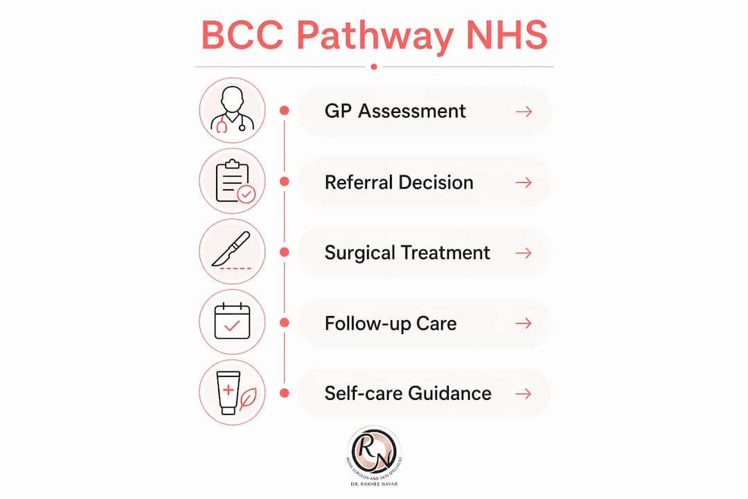
Basal cell carcinoma (BCC) is the most common skin cancer in the UK, accounting for roughly 75% of all skin cancer diagnoses. It grows slowly, rarely spreads to other organs, but causes significant local tissue damage and disfigurement if left untreated. The NHS manages BCC through a structured pathway: GP assessment, biopsy confirmation, surgical excision, and primary care follow-up. Understanding how the BCC NHS pathway works helps you act quickly, ask the right questions, and make informed decisions about your care.
How is BCC diagnosed and referred within the NHS?
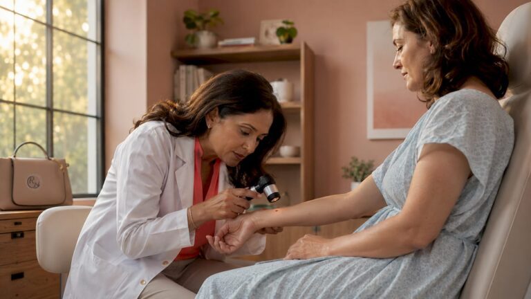
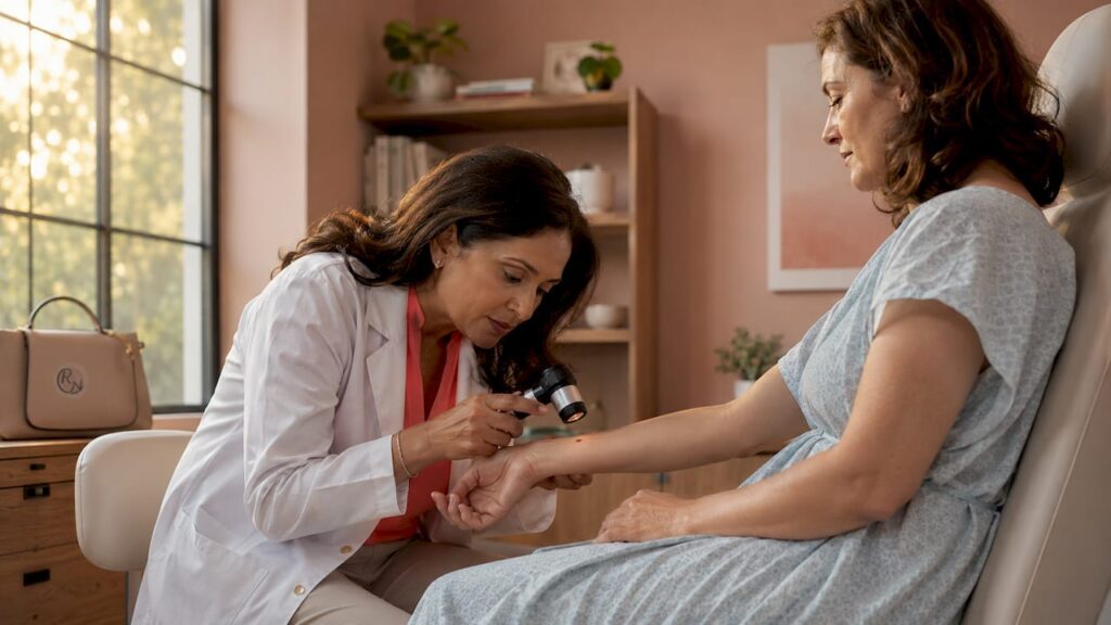
BCC diagnosis begins with your GP. A clinician examines the suspicious lesion and assesses features such as a pearly or translucent border, rolled edges, telangiectasia (visible small blood vessels), or central ulceration. These clinical signs are often enough to raise concern, but a biopsy and histopathological report remain the definitive method of confirmation.
Routine versus urgent referral
The NHS does not operate a national screening programme for basal cell carcinoma or other non-melanoma skin cancers. Cancer Research UK confirms this position is based on BCC’s slow growth rate and the effectiveness of patient self-identification. Routine referrals are processed within 18 weeks, which is appropriate for low-risk lesions on the trunk or limbs with no aggressive features.
High-risk lesions trigger a different response. BCCs located near the eyelid, ear, nose, or in proximity to major nerves or blood vessels require urgent specialist referral via NHS systems such as Sci Gateway in Scotland. These anatomical sites carry a higher risk of incomplete excision and functional impairment, so speed matters.
The histological subtype also influences urgency. Morphoeic (sclerosing) and infiltrative BCCs have poorly defined margins and a higher recurrence risk than nodular or superficial subtypes. A GP who identifies these features on biopsy will escalate the referral accordingly.
- Self-examination. Check your skin monthly, paying attention to new or changing lesions, particularly on sun-exposed areas such as the face, scalp, ears, and neck.
- GP consultation. Book an appointment promptly if a lesion bleeds without trauma, fails to heal, or changes in size or colour over four to six weeks.
- Biopsy. Your GP or dermatologist takes a small tissue sample under local anaesthetic. Results typically return within two to three weeks.
- Specialist referral. Depending on lesion risk, you are referred to dermatology or plastic surgery, either routinely or urgently.
Pro Tip: Photograph any suspicious lesion with your phone, including a ruler or coin for scale. A dated photo series helps your GP assess change over time and strengthens the case for referral.
What BCC treatment options does the NHS offer?
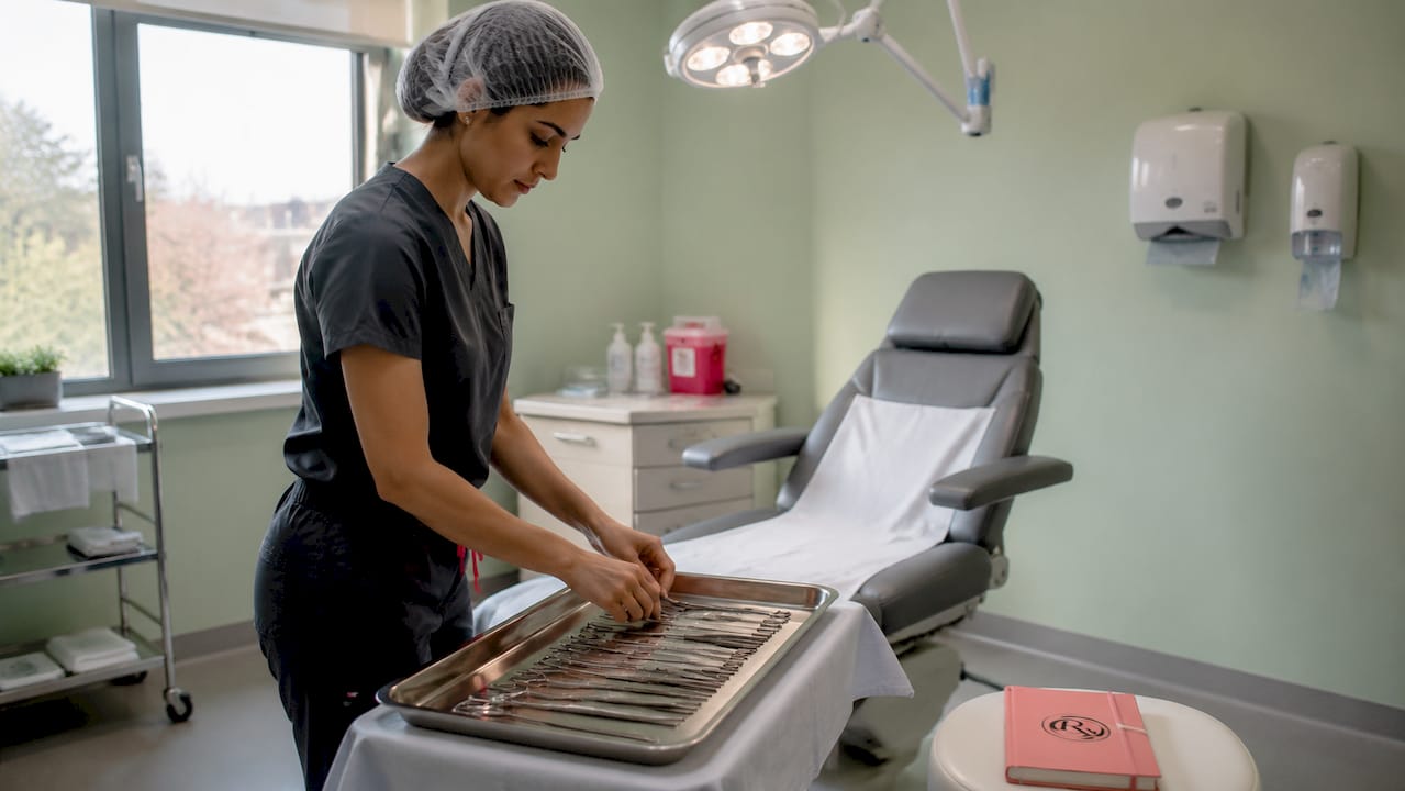
Surgical excision remains the cornerstone of BCC treatment within the NHS. The standard approach for low-risk lesions in primary care involves removing the tumour with a 4 mm lateral margin down to the subcutaneous fat. This margin is sufficient to achieve clear histological margins in the majority of well-defined, nodular BCCs on the trunk and limbs.
NHS treatment decisions are shaped by lesion location, size, histological subtype, and patient factors including age and comorbidities. A frail elderly patient with a small, low-risk BCC on the back may be managed differently from a younger patient with a morphoeic lesion on the nasal tip.
Non-surgical and topical treatments
Not every BCC requires a scalpel. The NHS offers several alternatives for selected patients.
- Imiquimod cream (Aldara). A topical immune response modifier applied to superficial BCCs, typically over six weeks. Suitable for thin, well-defined lesions on the trunk or limbs in patients unsuitable for surgery.
- Cryotherapy. Liquid nitrogen destroys superficial BCC tissue. Effective for small, low-risk lesions but not recommended for lesions near the eyes or on the face where margin control is critical.
- Radiotherapy. Used when surgery is not feasible due to patient health, lesion size, or anatomical complexity. Also used post-operatively if excision margins are involved and re-excision is not possible.
- Photodynamic therapy (PDT). Light-activated treatment for superficial BCCs, available in some NHS dermatology departments.
Advanced and systemic treatments
Vismodegib is approved by NHS England for locally advanced BCC as a neoadjuvant treatment prior to curative surgery, under strict clinical criteria. It targets the Hedgehog signalling pathway, which drives BCC cell growth. Sonidegib is a related agent used in similar circumstances. Both are reserved for adult patients with advanced or metastatic BCC who are not suitable for surgery or radiotherapy. These are not first-line options and carry significant side effects, including muscle cramps, hair loss, and dysgeusia (altered taste).
| Treatment | Best suited for | NHS availability |
|---|---|---|
| Surgical excision (4 mm margin) | Low-risk, well-defined BCCs on trunk/limbs | Widely available in primary and secondary care |
| Mohs micrographic surgery | High-risk, facial, or recurrent BCCs | Specialist centres; secondary/tertiary referral |
| Imiquimod cream | Superficial BCCs on trunk/limbs | Available on NHS prescription |
| Cryotherapy | Small, superficial, low-risk BCCs | Available in primary and secondary care |
| Radiotherapy | Inoperable or post-operative margin involvement | Available in NHS oncology departments |
| Vismodegib / sonidegib | Advanced or metastatic BCC; not suitable for surgery | NHS England approved under strict criteria |
Pro Tip: Ask your clinician specifically about the histological subtype of your BCC. Nodular and superficial subtypes respond well to standard excision. Morphoeic and infiltrative subtypes often need Mohs surgery or wider margins to achieve clearance.
How does NHS follow-up work after BCC treatment?
Most patients are discharged back to primary care after successful BCC excision. Intensive hospital follow-up is not standard NHS practice for low-risk, completely excised BCCs. This surprises many patients who expect regular dermatology appointments for years after treatment. The NHS model places responsibility for ongoing monitoring with the GP and the patient.
The rationale is sound. Patients who have had one BCC carry an estimated 50% risk of developing a further skin cancer within five years. That figure underlines why self-examination and sun protection are not optional extras. They are the primary tools for catching new lesions early.
What to watch for after treatment
The following signs warrant prompt re-consultation with your GP or specialist.
- A new lesion appearing anywhere on the skin, particularly on sun-exposed areas.
- Any change in the appearance of the treated site, including redness, swelling, or a new lump within or around the scar.
- A lesion that bleeds spontaneously or fails to heal within four weeks.
- Numbness, tingling, or altered sensation near a previously treated site, which may indicate nerve involvement.
Sun protection and self-care
The NHS advises daily broad-spectrum sunscreen (SPF 30 or higher), protective clothing, and avoidance of peak UV hours between 11am and 3pm. These measures reduce the risk of new BCCs forming. Tanning beds are a recognised risk factor and should be avoided entirely.
Genetic evaluation for multiple BCCs
Individuals who develop more than five BCCs before the age of 50 may be referred for genetic evaluation for Gorlin syndrome, also known as Nevoid Basal Cell Carcinoma Syndrome. This inherited condition predisposes individuals to multiple BCCs, jaw cysts, and other tumours. NHS genetics services, including those provided by South East Genomics, assess eligibility for testing based on clinical criteria. Early identification allows for more frequent surveillance and targeted management.
For detailed guidance on post-treatment skin checks, the follow-up steps outlined by Rakhee Nayar – Mohs Surgeon and Skin Specialist provide a practical framework for patients returning to primary care.
Standard excision vs Mohs surgery: what is the difference?
Standard surgical excision and Mohs micrographic surgery both remove BCC effectively, but they serve different clinical situations. Understanding the distinction helps you ask informed questions at your NHS appointment.
Standard excision removes the tumour with a predetermined margin, typically 4 mm for low-risk lesions. The specimen is sent to a laboratory and results return within one to two weeks. If the margins are involved, a second operation is required. This approach is appropriate for the majority of BCCs: well-defined, nodular or superficial lesions on the trunk, limbs, or non-critical facial areas.
Mohs micrographic surgery provides superior margin control and tissue preservation for high-risk or cosmetically sensitive lesions. The surgeon removes the tumour in thin layers, examining 100% of the surgical margin under the microscope in real time. If cancer cells remain at the edge, only the affected area is removed in the next layer. This continues until the margin is clear. The result is the smallest possible wound with the highest possible certainty of complete removal.
| Feature | Standard excision | Mohs micrographic surgery |
|---|---|---|
| Margin examination | Representative sections (approx. 1%) | 100% of surgical margin |
| Same-day result | No (1–2 weeks) | Yes |
| Tissue preservation | Standard | Maximum |
| Best suited for | Low-risk BCCs; trunk and limbs | High-risk, facial, recurrent BCCs |
| NHS availability | Widely available | Specialist centres |
| Recurrence rate (primary BCC) | Low for low-risk lesions | Lowest available for high-risk lesions |
The NHS directs patients with high-risk BCCs near the eyelid, nose, ear, or lip to specialist centres where Mohs surgery is available. Recurrent BCCs, those with aggressive histological subtypes, and lesions in areas where tissue preservation is critical also meet NHS criteria for Mohs referral.
Pro Tip: If your BCC is on your face and your GP has not mentioned Mohs surgery, ask specifically whether you meet the criteria for specialist referral. You are entitled to request this discussion.
For a detailed comparison of both approaches, the guide on Mohs vs standard excision from Rakhee Nayar – Mohs Surgeon and Skin Specialist sets out the clinical considerations clearly.
Key takeaways
The NHS manages BCC through a clear pathway: early GP assessment, biopsy confirmation, surgical excision with appropriate margins, and primary care follow-up, with specialist referral for high-risk or facial lesions.
| Point | Details |
|---|---|
| BCC prevalence | BCC accounts for roughly 75% of all UK skin cancer diagnoses, making it the most common type managed by the NHS. |
| No national screening | The NHS has no BCC screening programme; patient self-examination and GP vigilance are the primary detection tools. |
| Standard excision margin | A 4 mm lateral margin to fat depth is the NHS standard for low-risk BCC excision in primary care. |
| Follow-up responsibility | Most patients return to primary care after treatment; the 50% five-year risk of further skin cancers makes self-monitoring non-negotiable. |
| Mohs surgery criteria | High-risk, facial, or recurrent BCCs meet NHS criteria for Mohs micrographic surgery at specialist centres. |
What I have learned treating BCC patients in the NHS and beyond
Miss Rakhee Nayar shares her perspective below.
The single most common concern I hear from patients is not about the surgery itself. It is about what happens afterwards. People leave their NHS appointment with a healed scar and a letter discharging them to their GP, and they feel abandoned. They expected years of hospital check-ups. What they got was a leaflet about sunscreen.
I want to be direct about this: the NHS model is clinically sound. For a completely excised, low-risk BCC, intensive hospital surveillance adds little value. The evidence does not support it. What the evidence does support is that patients who understand their own risk, who check their skin monthly, and who return to their GP promptly when something changes, do well. The system works when patients are active participants in it.
Where I see the NHS pathway fall short is in the grey zone: lesions that are borderline for Mohs criteria, patients who are not told their histological subtype, and referrals that go to general surgery rather than a specialist with dual training in oncology and reconstruction. A morphoeic BCC on the nasal ala is not the same clinical problem as a nodular BCC on the back. Treating them identically is a mistake that leads to recurrence and more complex surgery later.
My advice to any patient reading this: know your subtype, know your site, and do not accept a standard excision for a high-risk facial lesion without asking whether Mohs surgery is appropriate. The NHS has the pathway. You need to know how to access it.
Lifelong sun protection is not a lifestyle suggestion. After one BCC, your skin has demonstrated its vulnerability. SPF 50, protective clothing, and avoiding midday sun are the most effective tools you have for preventing the next lesion.
— Miss Rakhee Nayar
Expert BCC care beyond the NHS: how Rakhee Nayar – Mohs Surgeon and Skin Specialist can help

For patients who want specialist assessment outside NHS waiting times, or whose BCC sits in a cosmetically sensitive area requiring both oncological precision and reconstructive expertise, Rakhee Nayar – Mohs Surgeon and Skin Specialist offers private consultations at Circle Cheshire in North West England. Miss Nayar holds dual training in plastic surgery and Mohs micrographic surgery, a combination that is rare in the UK and directly relevant to complex facial BCCs. Private consultations, e-consultations, and in-person assessments are available for UK and international patients. To understand your options, begin with the skin cancer symptoms guide or explore the full Mohs surgery service for detailed information on what to expect.
This article is for informational purposes only and does not constitute medical advice. Consult a GMC-registered specialist for assessment and treatment recommendations specific to your situation.
FAQ
What is BCC and how does the NHS treat it?
Basal cell carcinoma is the most common skin cancer in the UK, accounting for approximately 75% of all skin cancer cases. The NHS treats it primarily through surgical excision, with specialist options including Mohs micrographic surgery for high-risk or facial lesions.
Is there NHS screening for basal cell carcinoma?
No. The NHS does not operate a national BCC screening programme. Patients are advised to examine their own skin regularly and consult their GP promptly if a lesion changes, bleeds, or fails to heal.
When does the NHS refer a BCC patient urgently?
BCCs near the eyelid, ear, nose, or major nerves or blood vessels trigger urgent specialist referral. Aggressive histological subtypes such as morphoeic or infiltrative BCC also warrant expedited assessment due to their higher recurrence risk.
What is the risk of getting another BCC after treatment?
Patients who have had one BCC carry an estimated 50% risk of developing a further skin cancer within five years. Regular self-examination and daily sun protection are the most effective measures for reducing this risk.
When is Mohs surgery used instead of standard excision on the NHS?
Mohs micrographic surgery is recommended for BCCs on the face, particularly near the eyelid, nose, ear, or lip, and for recurrent or histologically aggressive lesions. It offers complete margin examination in real time, reducing recurrence risk and preserving healthy tissue.