
TL;DR:
- Most melanoma cases develop as completely new skin marks, not from existing moles. Early detection through structured checklists and regular self-examinations significantly improves treatment outcomes. Prompt medical evaluation of suspicious signs, especially non-healing sores or rapid growth, is crucial for effective skin cancer management.
Most people assume skin cancer announces itself through a mole they have had for years. In reality, 70% of melanoma cases form as entirely new marks on the skin, not changes to familiar moles. This single fact is why understanding skin cancer symptoms and signs matters far more than simply watching old spots. In the UK, skin cancer is the most common cancer overall, yet it carries an excellent prognosis when caught early. Knowing what to look for, where to look, and when to act is the difference between a minor surgical procedure and a much more involved course of treatment.
Key Takeaways
| Point | Details |
|---|---|
| Early detection saves lives | Most skin cancers can be cured if found and treated early through regular self-checks. |
| New marks matter | About 70% of melanomas start as new marks, so monitor your skin regularly for anything new. |
| Use the UK checklist | The weighted 7-point checklist helps healthcare professionals identify suspicious pigmented lesions needing urgent referral. |
| Urgent symptoms to note | Non-healing sores, bleeding moles, and rapidly growing lesions require immediate medical attention. |
| Seek expert care promptly | Prompt GP assessment and specialist referral mean skin cancer is more often found at an early, treatable stage. |
Common types of skin cancer and how their symptoms differ
Recognising skin cancer symptoms and signs starts with understanding that not all skin cancers look the same. The three main types seen in the UK, including melanoma, basal cell carcinoma (BCC), and squamous cell carcinoma (SCC), each present differently. Knowing their individual characteristics is the first step in using any skin cancer warning signs checklist effectively.
Melanoma is the most serious type. It often appears as a new mark rather than a change to an existing mole. It can be asymmetric, with irregular borders and more than one colour, ranging from tan and brown to black, pink, or even white. Some melanomas are flat; others are raised. A small number are amelanotic, meaning they contain no pigment at all and appear as a pale, skin-coloured, or faintly pink patch.
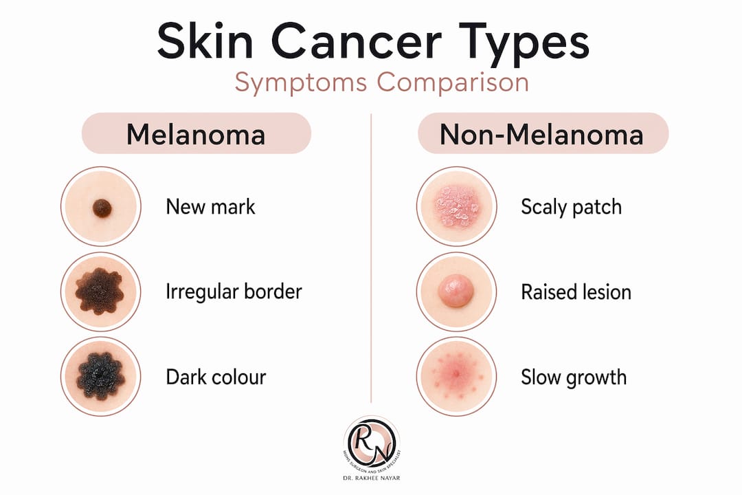
Basal cell carcinoma is the most common skin cancer in the UK. It usually appears on the face, ears, or neck, areas with a history of sun exposure. BCC typically presents as a raised, smooth, shiny, pearly bump with visible blood vessels running across its surface. It may also look like a flat, scar-like lesion or a pinkish patch with a slightly rolled edge. Many people describe early BCCs as a spot that simply refuses to heal.
Squamous cell carcinoma tends to look rougher and more obviously abnormal. SCC appears as scaly or crusty raised lesions, sometimes with a bleeding ulcer at the centre. The surrounding skin may look inflamed or thickened. SCC can grow faster than BCC and carries a greater risk of spreading if left untreated.
Here is a quick comparison of key features across the three types:
| Feature | Melanoma | Basal cell carcinoma | Squamous cell carcinoma |
|---|---|---|---|
| Appearance | New or changing mark, irregular colour | Pearly, shiny bump, rolled edges | Scaly, crusty, raised lesion |
| Common locations | Anywhere on the body | Face, ears, neck, scalp | Sun-exposed areas, lips, ears |
| Texture | Flat or raised | Smooth or waxy | Rough, hard, ulcerated |
| Bleeding | Possible, spontaneous | Occasional | Common, especially with pressure |
| Growth speed | Variable, can be rapid | Slow | Moderate to fast |
For a fuller overview, the types of skin cancer page provides detailed visual and clinical descriptions. You can also read about basal cell carcinoma and squamous cell carcinoma in depth.
Recognising melanoma signs using the UK clinical checklist
With an understanding of melanoma’s clinical indicators, you can use practical tools to assess suspicious skin changes. In the UK, GPs use the weighted 7-point checklist to evaluate pigmented lesions systematically, rather than relying on instinct alone.

The checklist divides features into major and minor signs, each carrying a different numerical weight:
Major features (2 points each):
- Change in size of a mole or new lesion
- Irregular shape or border
- Irregular colour (patchy, multi-toned)
Minor features (1 point each):
4. Largest diameter 7mm or more
5. Inflammation or redness around the lesion
6. Oozing, crusting, or bleeding
7. Change in sensation, such as itching or pain
A score of 3 or above triggers an urgent suspected cancer pathway referral to a dermatologist. This is not a tool for self-diagnosis. It is a guide that helps both patients and GPs recognise which lesions need fast-track assessment.
This system exists precisely because the human eye, without training, misses a significant proportion of early melanomas. A lesion that scores only 2 points may still warrant a GP review, particularly if it is new, fast-growing, or if you have a personal or family history of skin cancer.
Pro Tip: Photograph any suspicious mark with your phone. Date the image and take another in 4 to 6 weeks. A comparison photograph is one of the most useful pieces of information you can bring to a GP appointment.
For more detail on how lesions are assessed, see skin cancer detection and guidance on early skin cancer detection in the UK.
How to safely check your skin at home for cancer signs
Now that you know what signs to look for, here is how to perform an effective self-examination. The British Skin Foundation recommends checking your skin monthly. Since about 70% of melanomas are new marks, you are not simply keeping an eye on familiar moles. You are scanning your entire skin for anything unfamiliar.
What to do during a skin self-check:
- Use a well-lit room with a full-length mirror. Use a hand mirror for the back of your scalp, neck, back, and behind the ears.
- Check less visible areas including the soles of your feet, between your toes, under your nails, your groin, and the skin between your buttocks.
- Examine your lips, inside your mouth, and your genitals. Skin cancer can appear anywhere.
- Look for marks that are new, growing, changing in colour or shape, bleeding without being knocked, persistently itching, or failing to heal within a few weeks.
- Do not only look for dark or pigmented marks. BCCs and some melanomas are pale or skin-coloured.
“Any new or changing mark that bleeds, itches, or persists beyond a few weeks should be reported to your GP promptly. Do not wait to see if it resolves on its own.”
Pro Tip: Many UK GPs offer telephone or online appointments for skin concerns. If you are uncertain about a mark, take a photograph and contact your surgery. You do not need to be certain before reporting it.
If you have recently undergone treatment for skin cancer, follow-up skin care guidance will help you understand what to watch for during recovery.
Other high-urgency skin cancer symptoms to watch for
Beyond common mole changes, there are additional skin cancer warning signs that require immediate medical advice. These are the signs that fall outside the usual mole-monitoring framework, which means they are more likely to be overlooked.
- Non-healing sores: A wound, spot, or ulcer that fails to heal within three weeks, even if it scabs over and partially improves, can indicate basal cell carcinoma. The skin around it may look slightly shiny or pinkish.
- Spontaneous bleeding: A mole or mark that bleeds without being caught on clothing or scratched should be assessed without delay. Spontaneous bleeding and non-healing sores are among the highest-urgency warning signs a clinician would act on immediately.
- Rapidly growing nodules: A firm, red or pink nodule that appears and grows over weeks, particularly on sun-damaged skin, can be an SCC. These lesions sometimes look like an inflamed cyst. For more on early symptoms, the squamous cell carcinoma symptoms page is worth reviewing.
- Skin cancer in darker skin tones: Melanoma in people with darker skin often develops in locations with less sun exposure. This includes the palms of the hands, soles of the feet, under fingernails or toenails (appearing as a dark streak), and on mucous membranes. These presentations are frequently missed because they do not fit the stereotypical image of skin cancer.
Pro Tip: A dark streak running lengthwise under a nail is called subungual melanoma. It is rare, but it is also frequently diagnosed late because it is mistaken for bruising or a fungal infection. If the streak does not move as the nail grows, seek a medical opinion.
What happens after you report suspicious skin changes to your GP
Knowing when and how to seek expert help ensures you do not sit on a potentially significant skin change. Here is what the UK clinical pathway looks like once you make that first appointment.
- GP assessment: Your GP examines the lesion and applies the weighted 7-point checklist for pigmented marks, or uses clinical judgement for non-pigmented lesions such as BCCs. They will ask about changes over time, which is why that photograph you took is valuable.
- Fast-track referral: Suspected cancer pathway referrals are triggered for pigmented lesions scoring 3 or above, or for non-melanoma lesions with high-risk features. This pathway targets a specialist review within two weeks.
- Specialist examination: A dermatologist or skin cancer specialist assesses the lesion with a dermatoscope (a magnifying tool that reveals subsurface structures invisible to the naked eye) and decides whether a biopsy is required.
- Biopsy and diagnosis: A small sample of tissue is taken under local anaesthetic and sent to a pathologist. Results typically return within one to two weeks and confirm the type of cancer, its depth, and its margins.
- Treatment planning: Based on the biopsy results, the specialist recommends treatment. For higher-risk cancers or those in cosmetically sensitive areas such as the face, you may be referred to a surgeon with specialist skin cancer expertise. Details about the referral and treatment pathway can help you prepare for what to expect.
Why early detection and awareness save lives: a fresh look
Here is the uncomfortable truth about how most people actually approach skin cancer: they wait. They wait for a mole to look undeniably wrong. They wait for pain. They wait until the mark has been there so long it has become familiar. And familiarity, when it comes to skin cancer, is genuinely dangerous.
The assumption that skin cancer is always obvious is what makes it so frequently diagnosed late. People expect a suspicious lesion to look dramatic. Many do not. The most clinically significant skin changes are often quiet, slow, and easy to rationalise away.
What changes outcomes is structure. The reason the UK’s 7-point checklist works is not because it is sophisticated. It works because it replaces a subjective, emotionally loaded judgement (“is this serious?”) with a repeatable, objective scoring process. Structured clinical checklists reduce the delay caused by patient uncertainty and GP variability. They standardise the decision to refer, which means more cancers are caught while they are still highly treatable.
The same principle applies to self-checks. Monthly skin checks do not work because people suddenly become expert dermatologists. They work because they build a habit of looking, which means new marks get noticed sooner. Most skin cancers are curable if detected at an early stage. The British Skin Foundation’s emphasis on regular checks is not cautious box-ticking. It is grounded in genuine outcome data.
The lesson here is not to panic about every new freckle. It is to stop treating vigilance as optional. Monthly checks, prompt GP reporting, and understanding the clinical signs are not just good habits. They are the mechanism by which early-stage, highly curable skin cancer gets caught before it becomes something far more involved to treat. Read more about the value of early skin cancer detection and why timing changes everything.
Expert skin cancer diagnosis and treatment options in the UK
If you have noticed suspicious skin changes and want access to specialist-level assessment and treatment, the expertise available at mohssurgeon.co.uk is designed precisely for this.

Miss Rakhee Nayar holds dual training in plastic surgery and Mohs micrographic surgery, a combination that is rare in the UK and particularly valuable when treating skin cancers on the face, ears, and neck. Mohs micrographic surgery removes cancerous tissue layer by layer, with immediate microscopic examination to confirm clear margins. This method preserves as much healthy tissue as possible. It is the gold standard for BCCs and SCCs in cosmetically sensitive areas. Whether you need initial skin cancer detection services or are already managing a confirmed diagnosis, specialist input early in the process leads to better outcomes. You can explore high-risk skin cancer treatment options and book a private or e-consultation from anywhere in the UK.
Frequently asked questions
How often should I check my skin for signs of skin cancer?
Monthly skin checks are recommended to detect new or changing marks as early as possible, giving you the best chance of catching any concerning changes before they progress.
What should prompt me to see my GP about a skin mark?
See your GP if a mole or mark is new, growing, or if it changes in shape, colour, or bleeds, itches, or crusts without obvious cause. Do not wait to see whether it settles.
Can skin cancers appear on areas not exposed to the sun?
Yes. Melanoma on darker skin frequently develops on palms, soles, under nails, or on mucous membranes, areas that receive little to no sun exposure.
What is the importance of the weighted 7-point checklist in the UK?
The checklist allows GPs to assess pigmented lesions systematically. A score of 3 or more triggers an urgent two-week referral to a dermatologist, reducing delays in diagnosis caused by clinical uncertainty.
Are all skin cancers painful or itchy?
No. Many early skin cancers cause no discomfort at all. However, persistent itching or pain at a lesion site is considered a minor clinical feature and should prompt a GP review regardless of how the mark looks.
Recommended
- Why early skin cancer detection is crucial for better outcomes
- Squamous cell carcinoma symptoms: Know the signs early
- How to identify basal cell carcinoma on the face: UK guide
- Types of skin cancer and advanced facial treatments UK
Filed under Spotting Skin Cancer


