Basal cell carcinoma (BCC) on the nose is the most common form of skin cancer affecting the face, appearing as a shiny, pearly bump or a non-healing sore that requires prompt specialist assessment. BCC accounts for approximately 80% of all skin cancer cases, with the nose being one of the most frequently affected sites due to its prominence and constant sun exposure. Left untreated, nasal BCC is locally destructive and can damage underlying cartilage and soft tissue, even though it rarely spreads to distant organs. Early diagnosis and precise surgical removal are the cornerstones of effective treatment, and the choice of technique directly affects both cure rates and the long-term appearance of your nose.
What are the signs of BCC on the nose?
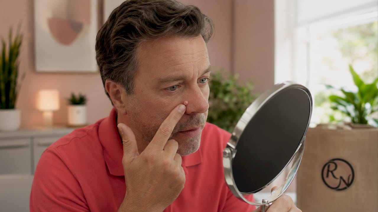
Recognising the early signs of basal cell carcinoma on the nose gives you the best chance of a straightforward treatment. The classic presentation is a small, shiny or pearly bump with a smooth, almost translucent surface. Many patients describe it as a spot that simply will not heal.
Common visual features
Typical nasal BCC lesions display one or more of the following characteristics:
- A pearly or waxy bump, often flesh-coloured or pale pink, sitting on the nasal skin or nasal tip
- Visible tiny blood vessels (telangiectasia) running across the surface of the lesion
- A non-healing sore that bleeds, crusts over, and then appears to partially heal before breaking down again
- A flat, scar-like patch that is slightly shiny and has poorly defined edges, more common with the morphoeic subtype
- Ulceration at the centre of the lesion, sometimes described as a rodent ulcer
Most nasal BCCs are painless in the early stages. Bleeding after minor trauma, such as rubbing the nose, is often the first symptom that prompts patients to seek advice. The morphoeic or infiltrative subtypes can be deceptively flat and pale, making them harder to spot without specialist examination.
Pro Tip: Take a clear photograph of any suspicious area on your nose in good natural light and note whether it has changed over four to six weeks. This simple record is genuinely useful when you attend a consultation.
Differentiating BCC from other conditions such as sebaceous hyperplasia, dermatofibroma, or squamous cell carcinoma requires clinical expertise. The signs of BCC on the face can overlap with benign lesions, which is why a professional assessment is always necessary before any treatment decision is made.
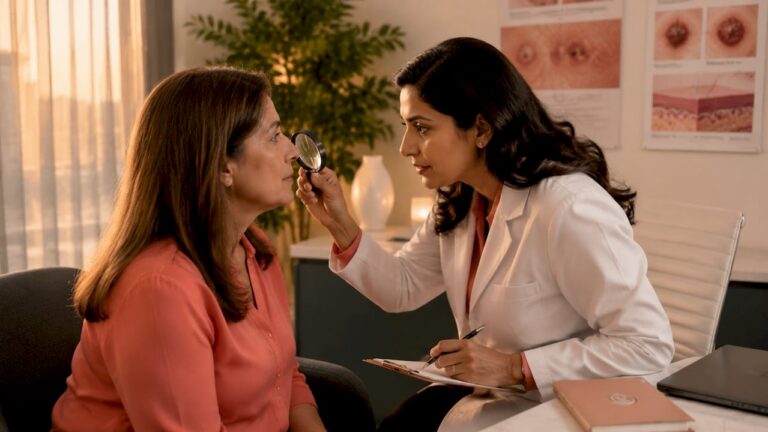
How is basal cell carcinoma on the nose diagnosed?
A clinical examination alone is not sufficient to confirm a diagnosis of nasal BCC. Biopsy and histological examination are mandatory for definitive diagnosis, particularly because mixed lesion types and rare variants can alter the treatment plan significantly. The diagnostic process typically follows a structured sequence.
The diagnostic pathway
- Clinical inspection — A consultant examines the lesion’s size, borders, colour, and surface texture. The location on the nose, whether the nasal tip, alar, sidewall, or dorsum, influences both the diagnosis and the reconstruction plan.
- Dermoscopy — A handheld dermatoscope magnifies the lesion and reveals subsurface features such as arborising blood vessels, blue-grey ovoid nests, and spoke-wheel structures that are characteristic of BCC.
- Incisional or punch biopsy — A small tissue sample is taken under local anaesthetic and sent to a histopathology laboratory. This confirms the diagnosis and identifies the BCC subtype, for example nodular, superficial, or morphoeic.
- Expert pathology review — Specialist pathological assessment is particularly important on the nose because morphoeic and infiltrative subtypes have indistinct margins that are easy to underestimate on standard excision.
- Staging and treatment planning — Once the subtype and extent are confirmed, the surgeon assesses whether the tumour is primary or recurrent, and whether imaging such as MRI is needed to evaluate deeper tissue involvement.
The distinction between a nodular BCC and a morphoeic BCC matters enormously for treatment planning. Nodular lesions tend to have well-defined edges and respond well to most surgical approaches. Morphoeic lesions spread silently through tissue planes and carry a higher recurrence risk if margins are not meticulously controlled.
What are the treatment options for BCC on the nose?
Treatment for BCC on the nose ranges from minor surgical procedures to specialist micrographic surgery and reconstructive plastic surgery. The right choice depends on the BCC subtype, tumour size, location on the nose, and whether the lesion is primary or recurrent.
Surgical excision
Standard surgical excision removes the tumour with a margin of surrounding normal tissue. Achieving clear surgical margins reduces the recurrence rate from approximately 24% with incomplete margins to around 6% with clear margins. This is a significant difference. The limitation of standard excision on the nose is that the surgeon examines only a small representative sample of the margins after the operation, not the entire margin in real time.
Mohs micrographic surgery
Mohs micrographic surgery is the gold standard for nasal BCC. The technique involves removing the tumour in thin layers and examining 100% of the surgical margins under the microscope while the patient waits. If any cancer cells remain at the edge, only that specific area is removed in the next layer. This continues until the margins are completely clear.
Mohs surgery achieves cure rates exceeding 99% for primary nasal BCC. That figure reflects the combination of complete margin control and the ability to spare healthy tissue. On the nose, where every millimetre of tissue matters for both function and appearance, this tissue-sparing property is particularly valuable.
Pro Tip: Ask your surgeon whether they are dual-trained in both Mohs surgery and plastic surgery. This combination means the same specialist who removes the cancer can also plan and perform the reconstruction, which leads to better-coordinated outcomes.
Reconstruction after tumour removal
Removing a BCC from the nose frequently leaves a defect that requires careful reconstruction. Effective nasal reconstruction depends on understanding the skin tension lines and nasal subunits, the anatomical zones of the nose such as the tip, alar, and dorsum, each of which has distinct skin thickness and texture. Reconstruction options include primary closure, local flaps such as the bilobed or nasolabial flap, skin grafts, and more complex forehead flaps for larger defects.
Non-surgical treatments
Non-surgical options including topical imiquimod, 5-fluorouracil cream, and photodynamic therapy (PDT) are suitable only for small, superficial BCCs. They carry lower cure rates than surgery and are not appropriate for nodular, morphoeic, or recurrent lesions on the nose. Radiotherapy is occasionally used in patients who are not fit for surgery, but it is not a first-line choice for nasal BCC given the long-term effects on nasal skin quality.
Treatment comparison
| Treatment | Cure rate | Tissue sparing | Best suited for |
|---|---|---|---|
| Mohs micrographic surgery | Over 99% (primary BCC) | Excellent | All nasal BCC, especially morphoeic, recurrent, or large lesions |
| Standard surgical excision | Around 94% with clear margins | Moderate | Well-defined, small primary nodular BCC |
| Curettage and electrodesiccation | Variable, lower than excision | Poor | Very small, superficial BCC only |
| Topical agents and PDT | Lower than surgical options | Not applicable | Small, superficial BCC in selected patients |
| Radiotherapy | Moderate | Poor long-term | Patients unfit for surgery |
The table makes clear that Mohs surgery for facial skin cancer consistently outperforms alternatives on the nose, where both cure and cosmetic outcome are priorities.
What does recovery involve after BCC removal from the nose?
Recovery after treatment for basal cell carcinoma on the nose depends on the extent of the surgery and the type of reconstruction performed. Most patients undergoing Mohs surgery with a straightforward closure or small flap return to normal activities within one to two weeks. Larger reconstructions involving forehead flaps require a staged approach and a longer recovery period.
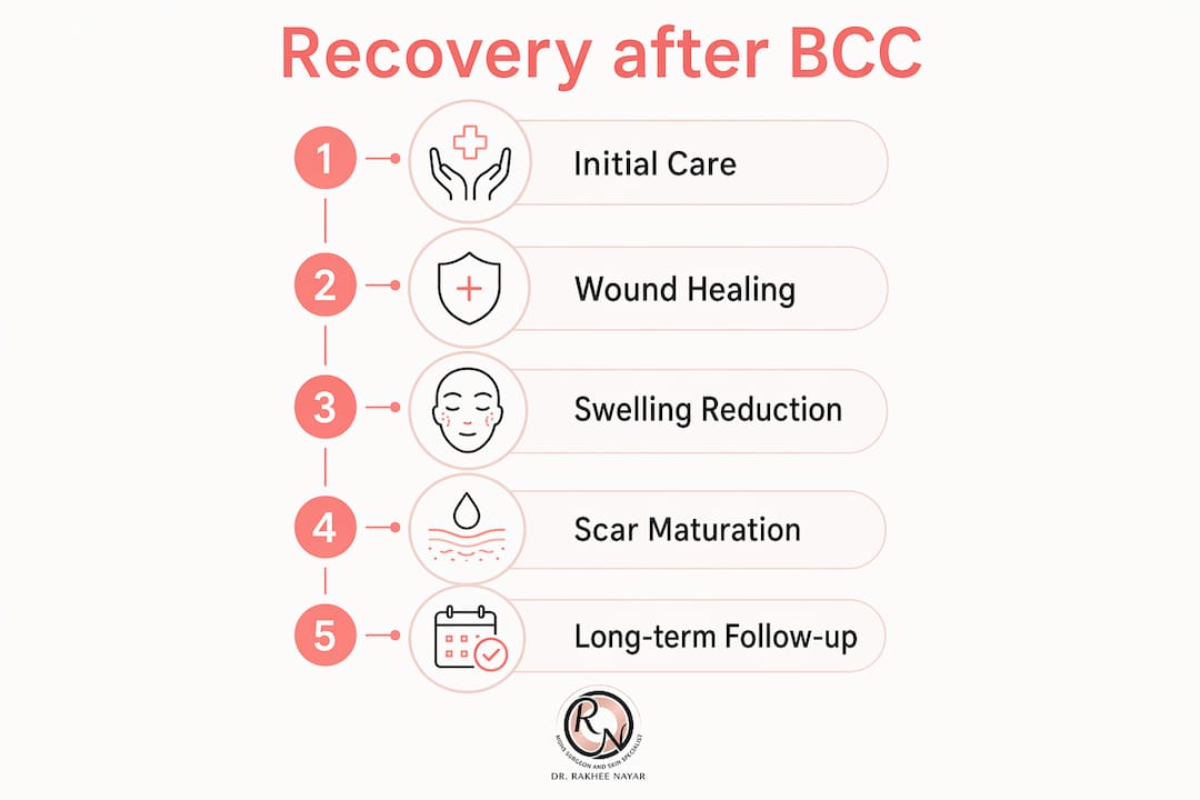
Wound care and healing
Structured post-operative care reduces the risk of infection, supports healing, and improves the final scar appearance. The key principles are:
- Keep the wound clean and moist with a thin layer of white soft paraffin or an antibiotic ointment as directed by your surgeon
- Avoid direct sun exposure on the healing wound for at least twelve months; use SPF 50+ sunscreen once the wound is fully closed
- Do not pick at crusts or scabs, as this disrupts the healing tissue and increases scarring
- Avoid strenuous activity for two to four weeks after surgery to reduce swelling and the risk of wound breakdown
- Attend all scheduled follow-up appointments, as early identification of wound complications or recurrence is far easier to manage than late presentation
Swelling around the nose is normal in the first one to two weeks and does not indicate a problem. Bruising may extend to the lower eyelids, particularly after surgery near the nasal bridge.
Monitoring for recurrence
The risk of BCC recurrence on the nose is highest in the first three years after treatment. Regular skin checks with a specialist are the most reliable way to detect any new or recurrent lesion early. Miss Nayar recommends a structured follow-up schedule, typically at three months, six months, and twelve months post-operatively, with annual skin checks thereafter.
Pro Tip: Perform a monthly self-examination of your nose and surrounding facial skin in a well-lit mirror. Look for any new shiny bump, persistent sore, or area of skin that looks different from the surrounding tissue. Report changes promptly rather than waiting for a scheduled appointment.
Skin protection and prevention
Sun protection is the single most effective strategy for reducing the risk of further skin cancers. Patients who have had one BCC on the nose have a meaningfully elevated risk of developing additional BCCs elsewhere on the face. Daily use of a broad-spectrum SPF 50+ sunscreen, protective clothing, and avoidance of peak UV hours between 11am and 3pm are all evidence-based measures. Vitamin D supplementation is worth discussing with your GP if you are significantly reducing sun exposure.
Key takeaways
Clear surgical margins are the strongest single predictor of successful treatment for nasal BCC, and Mohs micrographic surgery is the most reliable method for achieving them.
| Point | Details |
|---|---|
| BCC is the most common skin cancer | It accounts for approximately 80% of skin cancer cases and frequently affects the nose. |
| Early recognition matters | Pearly bumps, non-healing sores, and visible blood vessels on the nose warrant specialist review. |
| Biopsy confirms diagnosis | Clinical inspection alone is insufficient; histological examination identifies the BCC subtype and guides treatment. |
| Mohs surgery leads on cure rates | Cure rates exceed 99% for primary nasal BCC, with superior tissue preservation compared to standard excision. |
| Aftercare reduces recurrence risk | Structured wound care, sun protection, and regular follow-up are all required after BCC removal from the nose. |
Why nasal BCC deserves more respect than most patients give it
Patients often arrive at my clinic having been told by a well-meaning friend or a quick internet search that a BCC is “just a skin cancer” and nothing to worry about. I understand why that reassurance is offered. BCC rarely spreads to other organs, and the word “cancer” carries a weight that can feel disproportionate to a small shiny bump on the nose.
The reality I see in practice is more nuanced. BCC is locally destructive and, when it sits on the nose, it has the potential to invade cartilage, damage the nasal lining, and compromise the airway if left too long. I have treated patients who delayed seeking help for two or three years because the lesion seemed small and caused no pain. By the time they came to me, what might have been a straightforward Mohs procedure with a small flap had become a complex reconstruction involving multiple stages.
The other misconception I encounter regularly is that any surgeon can remove a BCC from the nose with equivalent results. Margin control is everything. The difference between a 6% recurrence rate with clear margins and a 24% rate with incomplete margins is not a minor statistical footnote. It represents a real patient who returns eighteen months later with a recurrent tumour that is now harder to treat and leaves a larger defect.
My approach is to treat every nasal BCC as a problem that deserves both precision and planning. That means choosing the right surgical technique for the subtype, understanding the nasal anatomy well enough to reconstruct it properly, and giving patients a clear picture of what recovery involves. Skin cancer on the nose is treatable with excellent outcomes. The key is not to underestimate it.
— Miss Rakhee Nayar
Expert Mohs surgery for nasal BCC at Rakhee Nayar – Mohs Surgeon and Skin Specialist
If you have noticed a suspicious lesion on your nose, or you have already received a BCC diagnosis and want to understand your options, Rakhee Nayar – Mohs Surgeon and Skin Specialist offers consultant-led assessment and treatment at Circle Cheshire in North West England.

Miss Nayar is dual-trained in Mohs micrographic surgery and plastic surgery, which means she combines precise tumour removal with expert nasal reconstruction in a single, coordinated care pathway. Whether you need a full Mohs surgery assessment or want to explore your facial reconstruction options, the clinic offers both in-person and e-consultations for UK and international patients. To book a private consultation or to learn more about the treatment pathway, visit mohssurgeon.co.uk.
This article is for educational purposes only and does not constitute medical advice. Please consult a GMC-registered specialist for assessment and treatment of any skin lesion.
FAQ
What does BCC on the nose look like?
Nasal BCC typically appears as a shiny, pearly or flesh-coloured bump, sometimes with visible tiny blood vessels on its surface. It may also present as a non-healing sore that bleeds, crusts, and partially heals repeatedly.
Is BCC on the nose serious?
BCC on the nose rarely spreads to other organs, but it is locally destructive and can damage cartilage and soft tissue if left untreated. Prompt treatment is important to prevent significant structural damage and to preserve nasal function and appearance.
What is the best treatment for BCC on the nose?
Mohs micrographic surgery is the recommended treatment for most nasal BCCs, offering cure rates exceeding 99% for primary tumours with superior tissue preservation. It is particularly suited to morphoeic, recurrent, or large lesions where margin control is critical.
How long does recovery take after BCC removal from the nose?
Recovery after straightforward Mohs surgery with a small flap typically takes one to two weeks for daily activities. Larger reconstructions may require a staged approach over several weeks, with full scar maturation taking up to twelve months.
Can BCC on the nose come back after treatment?
Recurrence is possible, particularly with incomplete surgical margins or aggressive subtypes. With clear margins achieved through Mohs surgery, the recurrence rate for primary nasal BCC is approximately 6% or lower, and regular follow-up appointments help detect any new lesion early.