Basal cell carcinoma (BCC) is defined as a malignant tumour arising from basal cells in the deepest layer of the epidermis, making it the most common form of skin cancer diagnosed in the United Kingdom. BCC is primarily caused by cumulative ultraviolet radiation exposure over many years. It grows slowly and invades local tissue but rarely spreads to distant organs. If you have recently received a BCC diagnosis, understanding what this means for your health, your treatment options, and your long-term skin care is the most productive place to start.
What is BCC skin cancer and why does it matter?
Basal cell carcinoma originates in the basal cells, which sit at the base of the outer skin layer and are responsible for producing new skin cells. Chronic sun exposure is the leading cause, though radiation therapy, arsenic exposure, and a weakened immune system also contribute. The condition accounts for the majority of all skin cancer diagnoses in the UK each year.
BCC is locally invasive, meaning it can destroy surrounding tissue, cartilage, and even bone if left untreated. This is the fact that surprises most newly diagnosed patients. The cancer rarely travels to lymph nodes or other organs, but delayed treatment can cause significant disfigurement and functional impairment, particularly on the face. Understanding this distinction between “rarely fatal” and “not serious” is critical to making informed decisions about your care.
You can read a detailed clinical overview of BCC’s features and diagnosis to supplement what your specialist has told you.
What are the symptoms and appearance of BCC?
BCC typically appears as a slowly enlarging, painless lesion that may look like a shiny, pearly, or translucent bump, often with visible blood vessels on its surface. Some lesions present as a flat, scaly, reddish patch that is easily mistaken for eczema or psoriasis. Others appear as a sore that bleeds, crusts over, and seems to heal, only to break down again.
Appearance varies considerably by skin tone. In people with lighter skin, the classic presentation is a pearly or flesh-coloured nodule, often on the nose, cheek, or forehead. In people with darker skin, the lesion may appear brown or glossy black, which can delay recognition. The face, ears, neck, and scalp account for the majority of cases, though BCC can occur anywhere on the body that has received sun exposure over a lifetime.
Common presentations to look out for include:
- A shiny, skin-coloured or pink bump with a translucent quality
- A flat, brown or flesh-coloured scar-like lesion
- A bleeding or scabbing sore that recurs after apparent healing
- A pink growth with raised edges and a crusted centre
- A white, waxy lesion with poorly defined borders (associated with morpheaform subtype)
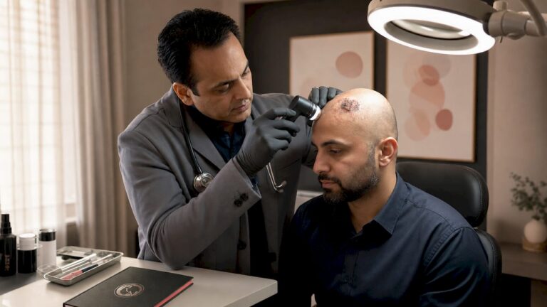
Pro Tip: If a lesion on your face or neck has bled more than once without an obvious cause, do not wait for it to resolve on its own. A biopsy is the only way to confirm or exclude BCC, and early confirmation changes your treatment options significantly.
Visual examples of BCC presentations can help you compare what you have noticed on your own skin with documented clinical cases.
How is basal cell carcinoma staged and classified?
BCC staging is less formalised than for cancers such as melanoma or breast cancer. Clinicians instead use a risk stratification system that divides lesions into low-risk and high-risk categories based on several clinical and histological factors. This distinction directly determines which treatment is appropriate for you.
More than 90% of BCCs are classified as low-risk. High-risk lesions are less common but require more precise management, often in specialist hands.
| Risk Factor | Low-Risk BCC | High-Risk BCC |
|---|---|---|
| Tumour size | Less than 6 mm (H-zone) or less than 10 mm (cheeks, forehead) | Greater than 6 mm (H-zone) or greater than 20 mm (trunk) |
| Location | Trunk, limbs, low-risk facial zones | H-zone (nose, ears, periocular, lips) |
| Border definition | Well-defined | Poorly defined or infiltrative |
| Histological subtype | Nodular, superficial | Morpheaform, infiltrative, micronodular |
| Prior treatment | Primary (untreated) | Recurrent after previous treatment |
| Perineural or vascular involvement | Absent | Present |
The H-zone of the face (nose, inner corners of the eyes, ears, and lips) is considered particularly high-risk because tumours in these areas can invade deeply into underlying structures with minimal surface warning. A lesion on the nose tip, for example, may extend to nasal cartilage before it becomes clinically apparent.
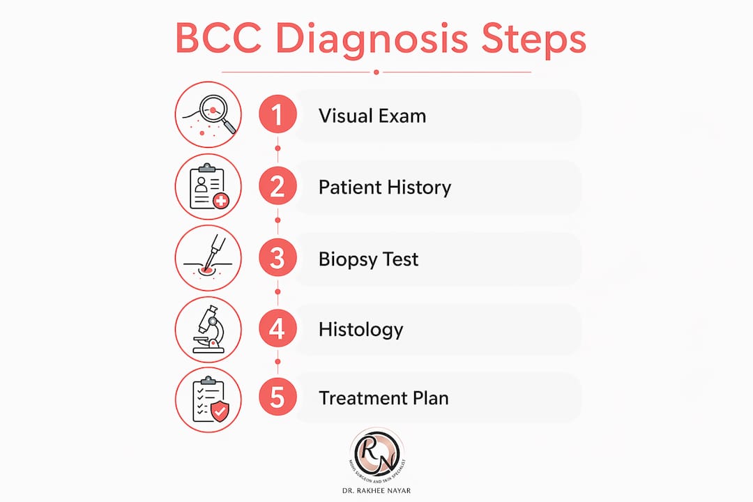
Biopsy is the only reliable method for confirming the diagnosis and identifying the histological subtype. Visual examination alone cannot determine whether a lesion is nodular (the most common and most treatable subtype) or morpheaform (a subtype with invisible margins that carries a higher recurrence risk). Knowing the subtype before treatment is not a formality. It is the information that guides every subsequent clinical decision.
What are the standard treatments for BCC?
Treatment for BCC depends on the lesion’s risk category, its histological subtype, its location, and your overall health. The goal in every case is complete removal with the lowest possible risk of recurrence and the best achievable cosmetic and functional outcome.
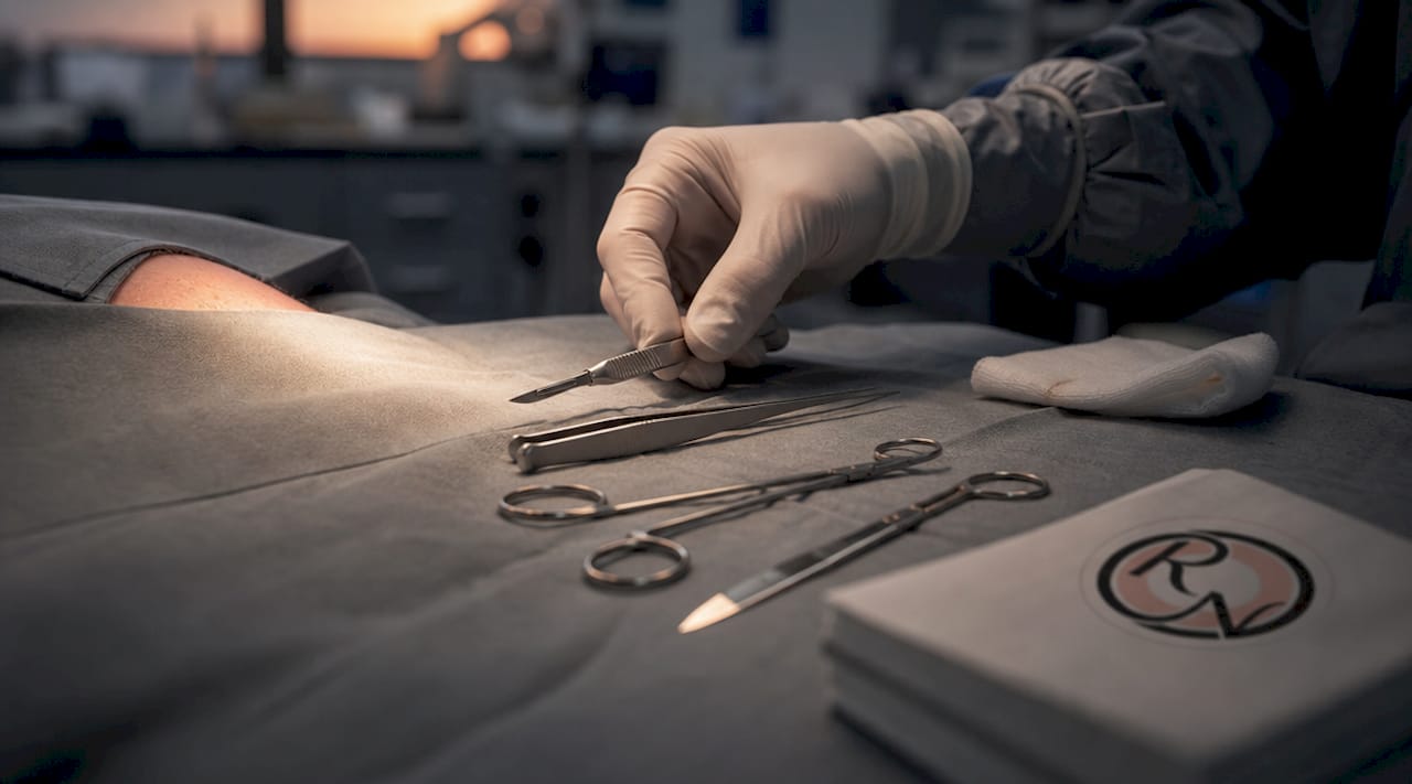
Surgical excision
Standard surgical excision is appropriate for most low-risk BCCs on the trunk and limbs. The surgeon removes the tumour with a margin of normal-appearing tissue around it, and the specimen is sent to a laboratory for margin assessment. This is a straightforward, well-established procedure performed under local anaesthetic.
Mohs micrographic surgery
Mohs micrographic surgery is the treatment of choice for high-risk BCCs, recurrent lesions, and tumours in cosmetically or functionally sensitive areas. The procedure involves removing the tumour in thin layers, examining 100% of the surgical margin under the microscope in real time, and continuing only until all margins are clear. This tissue-sparing approach achieves high cure rates while preserving as much healthy skin as possible.
Morpheaform and infiltrative subtypes have margins invisible to the naked eye, making standard excision unreliable for these cases. Mohs surgery was specifically designed to address this problem. For lesions on the nose, eyelids, lips, or ears, the functional and aesthetic stakes are high enough that tissue preservation is not optional. It is clinically necessary.
Pro Tip: Ask your surgeon specifically whether your BCC subtype has been confirmed by biopsy before agreeing to a treatment plan. The subtype determines whether standard excision is sufficient or whether Mohs surgery is the more appropriate choice.
Non-surgical options
Non-surgical treatments are available for selected low-risk, superficial BCCs where surgery is not suitable. These include:
| Treatment | Best Suited For | Limitations |
|---|---|---|
| Topical imiquimod (Aldara) | Superficial BCC on trunk or limbs | Not suitable for nodular or high-risk subtypes |
| Topical fluorouracil (5-FU) | Superficial BCC in low-risk locations | Lower cure rates than surgery |
| Cryotherapy | Small, superficial, well-defined lesions | No margin assessment; higher recurrence risk |
| Photodynamic therapy (PDT) | Superficial BCC, particularly in elderly patients | Not appropriate for deep or infiltrative lesions |
| Radiotherapy | Patients unfit for surgery | No tissue for histological margin assessment |
For advanced or metastatic BCC, which is rare, targeted therapies such as vismodegib (a hedgehog pathway inhibitor) are available. These are systemic treatments used when surgery and radiotherapy are no longer viable options.
The choice between Mohs surgery and standard excision is not simply a matter of preference. It is a clinical decision based on your specific lesion characteristics, and it should be made with a surgeon who has experience in both techniques.
How can you prevent BCC and what follow-up is needed?
Sun protection and regular skin examinations are the two most evidence-supported strategies for reducing BCC risk and catching new lesions early. Once you have had one BCC, your lifetime risk of developing another is significantly elevated. Prevention and monitoring are therefore ongoing commitments, not one-off actions.
Practical steps to reduce your risk include:
- Apply a broad-spectrum sunscreen of SPF 30 or higher every day, including in winter and on overcast days
- Wear protective clothing such as long sleeves, wide-brimmed hats, and UV-blocking sunglasses
- Avoid direct sun exposure between 11 am and 3 pm, when UV radiation is strongest
- Never use sunbeds or tanning lamps
- Perform a monthly self-examination of your skin, including the scalp, ears, and back of the neck
- Attend all follow-up appointments with your specialist, typically every 6–12 months after treatment
Follow-up care after BCC treatment serves two purposes. The first is to monitor the treated site for signs of recurrence. A lesion that appears healed on the surface can still harbour cancerous root structures underneath, which is why clinical review is not optional. The second purpose is to detect new primary BCCs early, when treatment is simpler and outcomes are better.
If you notice any new or changing lesion between scheduled appointments, contact your specialist promptly rather than waiting for your next routine review. Early detection consistently leads to less extensive surgery and better cosmetic results.
Key takeaways
Basal cell carcinoma is a locally invasive skin cancer that is highly treatable when diagnosed early and managed according to its histological subtype and risk category.
| Point | Details |
|---|---|
| BCC is the most common skin cancer | It arises from basal cells and is primarily driven by cumulative UV exposure over years. |
| Biopsy confirms subtype and guides treatment | Visual diagnosis alone is insufficient; subtype determines whether standard excision or Mohs surgery is appropriate. |
| High-risk lesions need specialist care | H-zone location, poor border definition, and aggressive subtypes require tissue-sparing Mohs surgery. |
| Local invasion is the primary risk | BCC rarely spreads to other organs but can destroy tissue, cartilage, and bone without timely treatment. |
| Prevention and follow-up are lifelong | Daily sun protection and regular specialist review reduce recurrence and catch new lesions at an earlier stage. |
What i have learnt from treating BCC every week
The single most common misconception I encounter in clinic is that a BCC diagnosis is “nothing to worry about.” Patients arrive having been told by a well-meaning friend or a brief internet search that it is “just a skin cancer” and that it will be easily removed. That framing is partially true and largely unhelpful.
BCC is treatable. In the vast majority of cases, it is curable with the right intervention. But the word “just” does a disservice to patients who then delay seeking specialist assessment, or who accept a treatment plan without asking whether it is the most appropriate one for their specific lesion. I have seen BCCs on the nose and inner canthus of the eye that have been inadequately excised elsewhere, only to recur with deeper invasion into cartilage or periorbital tissue. Recurrent BCC is harder to treat, requires more extensive surgery, and carries a greater risk of functional impairment.
The other thing I want patients to understand is that not all BCCs are the same. A superficial BCC on the back of a 70-year-old is a very different clinical problem from a morpheaform BCC on the nasal tip of a 55-year-old. The first may be managed with topical therapy or a simple excision. The second requires Mohs surgery performed by someone trained to examine margins intraoperatively and reconstruct the defect with both function and appearance in mind.
My advice to anyone newly diagnosed is this: ask for your biopsy result in writing, confirm the histological subtype, and ask your surgeon directly whether they have considered Mohs surgery for your lesion. If the answer is that it was not considered because the lesion is “straightforward,” ask why. You are entitled to that conversation. The seriousness of a BCC is not fixed. It depends on where it is, what type it is, and how it is treated.
— Miss Rakhee Nayar
Specialist BCC treatment at rakhee nayar – mohs surgeon and skin specialist
If you have been diagnosed with basal cell carcinoma and want a specialist opinion on your treatment options, Rakhee Nayar – Mohs Surgeon and Skin Specialist offers private consultations at Circle Cheshire in North West England, with e-consultations available for patients across the UK and internationally.

Miss Nayar holds dual training in plastic surgery and Mohs micrographic surgery, a combination that allows her to remove BCC with precise margin control and reconstruct the defect with the best possible cosmetic outcome. Whether your lesion is newly diagnosed or has recurred after previous treatment, a consultation will clarify your options. You can learn more about the Mohs surgery procedure or explore the step-by-step removal process before your appointment.
This article is for informational purposes only and does not constitute medical advice. Please consult a GMC-registered specialist for assessment and treatment recommendations specific to your situation.
FAQ
What is BCC skin cancer in simple terms?
Basal cell carcinoma is a type of skin cancer that grows from the basal cells at the base of the outer skin layer. It is the most common skin cancer in the UK, usually caused by long-term sun exposure, and it grows slowly without typically spreading to other parts of the body.
Is BCC skin cancer dangerous?
BCC is rarely life-threatening but can cause serious local damage if left untreated, including destruction of tissue, cartilage, and bone. Early treatment leads to straightforward outcomes; delayed treatment increases the risk of disfigurement and more extensive surgery.
How is BCC skin cancer diagnosed?
Biopsy is required to confirm the diagnosis and identify the histological subtype. Visual examination alone is not sufficient, as BCC presents differently across skin tones and subtypes, and the subtype directly determines which treatment is most appropriate.
What is the difference between BCC and melanoma?
BCC arises from basal cells and rarely metastasises, making it less immediately life-threatening than melanoma. Melanoma originates from pigment-producing melanocytes, spreads to lymph nodes and distant organs far more readily, and carries a significantly higher mortality risk. You can read more about skin cancer types and treatments to understand how they differ clinically.
Can BCC skin cancer come back after treatment?
Yes. Recurrence depends on the histological subtype, the completeness of the original excision, and the location of the lesion. Morpheaform and infiltrative subtypes carry the highest recurrence risk. Regular follow-up with a specialist and monthly self-examination are the most reliable ways to detect recurrence at an early, treatable stage.