Basal cell carcinoma (BCC) is the most common form of skin cancer, accounting for approximately 70% of all skin cancer cases in the UK. It arises from abnormal growth of basal cells, which sit at the deepest layer of the epidermis. BCC skin lesions typically appear on sun-exposed areas such as the face, scalp, ears, and neck, and they grow slowly over months or years. Most cases are curable when caught and treated early, but the right treatment depends heavily on the lesion’s size, location, and histological subtype.
What is BCC skin cancer and how does it develop?
Basal cell carcinoma is defined as a malignant tumour arising from basal keratinocytes in the lower epidermis. These cells normally divide to replenish the skin’s surface, but cumulative DNA damage causes them to multiply abnormally. The result is a locally invasive tumour that rarely spreads to other organs but can cause significant tissue destruction if left untreated.
BCC does not arise overnight. Most lesions represent decades of accumulated ultraviolet (UV) damage. This is why the condition is most common in adults over 50, though younger patients with high UV exposure histories are increasingly presenting with early lesions. The lifetime risk in the white UK population is estimated at 33–39% for men and 23–28% for women. That is a striking figure. It means roughly one in three men in the UK will develop a basal cell carcinoma at some point in their lifetime.
Understanding the biology matters because it directly shapes treatment. BCC lesions frequently extend microscopically beyond their visible borders, which is why surgeons plan excisions with wider margins than the lesion appears to require.
What causes basal cell carcinoma and who is most at risk?
UV exposure is the primary cause of basal cell carcinoma, both from natural sunlight and from artificial sources such as sunbeds. UV radiation damages the DNA in basal cells, particularly the tumour suppressor gene PTCH1, which normally regulates cell growth. Repeated damage over years overwhelms the skin’s repair mechanisms.
Certain individuals carry a significantly higher risk:
- Fair skin, light eyes, and red or blonde hair. People who burn easily and tan poorly accumulate UV damage more rapidly.
- Age over 50. Cumulative sun exposure increases with age, and the skin’s repair capacity declines.
- Outdoor occupations. Farmers, builders, gardeners, and sailors face prolonged daily UV exposure across their working lives.
- Immunosuppression. Organ transplant recipients on long-term immunosuppressive therapy have a substantially elevated risk of developing skin cancers, including BCC.
- Genetic syndromes. Gorlin syndrome (also known as basal cell naevus syndrome) causes multiple BCCs to develop from a young age, sometimes in the hundreds.
- Previous skin cancer. A prior BCC diagnosis is itself a significant risk factor for further lesions.
Sunbed use deserves specific mention. The British Skin Foundation and Cancer Research UK both identify sunbed use as a modifiable risk factor. Sunbeds emit UVA and UVB radiation at intensities that can exceed midday summer sunlight. Avoiding them entirely is the clearest preventive step available.
Pro Tip: If you work outdoors, apply a broad-spectrum SPF 50 sunscreen to exposed skin every morning, reapply every two hours, and wear a wide-brimmed hat. This is not optional skincare. It is primary prevention.

How to identify basal cell carcinoma on the skin
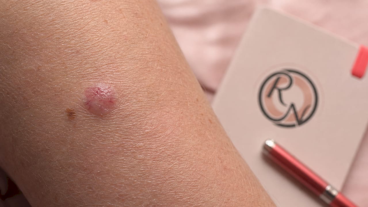
BCC lesions do not all look the same. Recognising the range of presentations is the first step toward seeking timely assessment. The most common appearance is a shiny, pearly, or translucent nodule with small visible blood vessels (telangiectasia) on the surface. However, BCC lesions may also present as red patches, scaly plaques, or ulcerated areas with rolled edges.
The four main subtypes each have distinct features:
- Nodular BCC. The most common subtype. A raised, dome-shaped, pearly lesion, often with a central depression or ulcer. Typically found on the nose, cheeks, or forehead.
- Superficial BCC. A flat, red, scaly patch that can resemble eczema or psoriasis. More common on the trunk and shoulders. Responds well to topical treatments.
- Morphoeic (sclerosing) BCC. A pale, scar-like plaque with indistinct borders. This subtype is the most difficult to treat because its true extent is rarely visible to the naked eye. Morphoeic and infiltrative subtypes are more aggressive and carry a higher recurrence risk.
- Basosquamous BCC. A mixed tumour with features of both BCC and squamous cell carcinoma. Considered higher risk due to greater metastatic potential than standard BCC.
Lesions commonly bleed after minor trauma, crust over, appear to heal, then bleed again. This cycle misleads many patients into thinking the lesion is resolving. Patients frequently delay treatment because of this deceptive healing pattern. A lesion that repeatedly scabs and bleeds over several weeks warrants medical review, not watchful waiting.
Pro Tip: Use the face identification guide to compare your lesion against clinical photographs before your GP appointment. Arriving with a clear description of the lesion’s behaviour and duration helps your clinician triage appropriately.
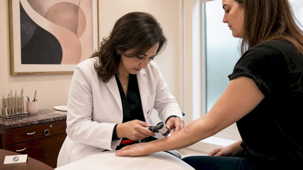
Diagnosis is confirmed by biopsy. A punch or excisional biopsy is preferred over a shave biopsy for suspected high-risk or recurrent BCC, as it provides sufficient tissue depth for accurate histological subtype assessment. The subtype identified on biopsy directly determines which treatment is most appropriate.
What are the main treatment options for BCC skin lesions?
Treatment selection depends on the lesion’s subtype, size, location, and whether it is a primary or recurrent tumour. No single approach suits every case. The table below summarises the main options and their typical outcomes.
| Treatment | Best Suited For | Approximate Cure Rate | Key Limitation |
|---|---|---|---|
| Surgical excision | Primary, low-risk nodular or superficial BCC | 95–98% | Requires adequate margins; may need reconstruction |
| Mohs micrographic surgery | High-risk, recurrent, facial, or morphoeic BCC | Over 99% (primary); 95% (recurrent) | Specialist availability; longer procedure |
| Topical 5-fluorouracil or imiquimod | Superficial BCC only | 80–95% | Higher recurrence than surgery; not for nodular types |
| Photodynamic therapy (PDT) | Superficial BCC, cosmetically sensitive sites | 80–90% | Multiple sessions; not suitable for thick lesions |
| Cryotherapy | Small, superficial, low-risk lesions | 85–95% | No histological confirmation; higher recurrence risk |
| Radiotherapy | Elderly or medically unfit patients | 90–95% | Multiple sessions; long-term skin changes |
| Hedgehog pathway inhibitors (vismodegib, sonidegib) | Advanced or metastatic BCC | Variable | Significant side effects; not a first-line option |
Surgical excision
Standard surgical excision with 3–5mm margins provides cure rates of 95–98% for primary BCC when appropriate margins are achieved. This is the most widely used treatment in the UK and is suitable for the majority of low-risk lesions on the trunk, limbs, and lower face. The procedure is performed under local anaesthetic as a day case. The excised tissue is sent to a pathology laboratory, and results confirming clear margins are typically available within one to two weeks.
Mohs micrographic surgery
Mohs surgery offers cure rates exceeding 99% for primary lesions and around 95% for recurrent tumours. It is the gold standard for BCCs on the face, ears, nose, eyelids, and lips, where tissue conservation is critical. The surgeon removes the tumour in thin layers, examining each layer under a microscope in real time before proceeding. This means no tissue is removed unnecessarily, and the wound is only closed once clear margins are confirmed. You can read a full step-by-step Mohs guide to understand what to expect on the day.
Non-surgical options
Topical treatments such as 5-fluorouracil and imiquimod achieve cure rates of approximately 80–95% for superficial BCC. These are applied at home over several weeks and are a reasonable option for patients with superficial lesions who are not surgical candidates. However, they carry higher recurrence rates than surgery and are not appropriate for nodular, morphoeic, or basosquamous subtypes.
Radiotherapy is suited to elderly or medically unfit patients who cannot tolerate surgery. Fractionated regimens over several weeks achieve control rates of around 90–95%. It is not a first choice for younger patients because long-term radiation changes to the skin can become cosmetically problematic over decades.
Pro Tip: Ask your clinician specifically about the histological subtype confirmed on biopsy before agreeing to a treatment plan. A superficial BCC on the back and a morphoeic BCC on the nose require very different approaches, even if they look similar on the surface.
What follow-up care is recommended after BCC treatment?
Follow-up after BCC treatment serves two purposes: detecting recurrence at the treated site and identifying new primary tumours elsewhere on the skin. Around 25% of patients diagnosed with BCC will develop a new primary tumour within five years. That figure underlines why follow-up is not optional.
The recommended follow-up schedule depends on risk:
- Low-risk primary BCC with clear margins. A single post-treatment review at three to six months is often sufficient. Patients are then discharged with self-surveillance advice.
- High-risk or recurrent BCC. Annual specialist review for at least five years is standard practice. Some patients with multiple lesions or Gorlin syndrome require indefinite monitoring.
- Immunosuppressed patients. More frequent review, often every three to six months, is warranted given the elevated risk of rapid new tumour development.
- Patients with multiple prior BCCs. A structured surveillance programme with a dermatologist or specialist nurse is appropriate.
Follow-up care is tailored by risk; self-surveillance and sun protection are critical between clinical appointments. Monthly self-examination is advised. Use a full-length mirror for the body and a hand mirror for the scalp and ears. Ask a partner or family member to check areas you cannot see clearly, particularly the back and the back of the neck.
Sun protection after treatment is not merely cosmetic. Daily use of SPF 50 broad-spectrum sunscreen, protective clothing, and avoidance of peak UV hours (11am to 3pm) reduces the risk of new lesions developing. This is the most evidence-based lifestyle modification available to BCC patients.
Pro Tip: Photograph any new or changing lesion on your skin with your smartphone and note the date. A series of dated photographs over four to six weeks gives your clinician far more useful information than a verbal description alone.
For detailed guidance on what to do after treatment, the follow-up steps after skin cancer treatment page covers surveillance schedules, self-examination technique, and when to seek urgent review.
What can patients expect during diagnosis and recovery?
Understanding what happens at each stage reduces anxiety and helps you make informed decisions. The process from suspicion to treatment completion typically spans several weeks.
At the biopsy stage:
- A punch or excisional biopsy is performed under local anaesthetic. The procedure takes around 15–20 minutes.
- The biopsy site heals within one to two weeks and leaves a small scar.
- Results confirming the subtype and grade are usually available within seven to ten working days.
- The subtype identified determines whether standard excision, Mohs surgery, or a non-surgical approach is most appropriate.
During surgical treatment:
- Most BCC procedures are performed under local anaesthetic as a day case. General anaesthetic is rarely required.
- Mohs surgery may take several hours if multiple layers are needed, but patients are awake and comfortable throughout.
- Wound closure depends on the defect size. Small wounds close directly. Larger defects may require a skin flap or graft, particularly on the nose, ear, or eyelid.
- Reconstructive surgery for facial lesions is an integral part of the procedure, not an afterthought. Miss Nayar’s dual training in plastic surgery and Mohs surgery means reconstruction is planned from the outset.
During recovery:
- Most patients return to normal activities within one to two weeks of standard excision.
- Mohs surgery wounds take slightly longer to heal, particularly if reconstruction was required.
- Scar maturation takes 12–18 months. Redness and firmness in the early months are normal and do not indicate a problem.
- Complications including infection, wound dehiscence, or haematoma are uncommon but should be reported promptly to your clinical team.
The psychological impact of a skin cancer diagnosis is often underestimated. Anxiety about recurrence, concern about scarring, and uncertainty about the future are all common. The British Association of Dermatologists and Macmillan Cancer Support both offer patient information resources and support networks for people living with skin cancer.
Key takeaways
Basal cell carcinoma is the most common skin cancer in the UK, and the right treatment, chosen according to lesion subtype and location, delivers cure rates above 95% in the majority of cases.
| Point | Details |
|---|---|
| BCC is the most common skin cancer | It accounts for approximately 70% of all skin cancer cases and carries a lifetime risk of up to 39% in UK men. |
| Subtype determines treatment | Nodular and superficial BCCs suit excision or topical therapy; morphoeic and recurrent lesions require Mohs surgery. |
| Mohs surgery offers the highest cure rate | Cure rates exceed 99% for primary lesions, making it the preferred choice for facial and high-risk BCCs. |
| New tumours are common after treatment | Around 25% of patients develop a new primary BCC within five years, making long-term self-surveillance critical. |
| Sun protection is primary prevention | Daily SPF 50 and protective clothing reduce the risk of new lesions and are recommended for all BCC patients post-treatment. |
What i have learned treating BCC over many years
The most consistent pattern I see in clinical practice is delay. Patients arrive having watched a lesion bleed, crust, and apparently heal for six, twelve, sometimes eighteen months before seeking assessment. The deceptive healing cycle of BCC is not a sign of recovery. It is the tumour’s normal behaviour. By the time some of these patients reach me, what began as a small nodule has become a lesion requiring complex reconstruction.
My view is that the conversation about BCC needs to shift. Too many people still think of skin cancer as something that only happens to people who sunbathe excessively. In reality, cumulative everyday UV exposure, the school run, the commute, the weekend gardening, is sufficient to cause significant damage over decades. Fair-skinned adults in their fifties and sixties who have spent their lives in the UK are at genuine risk, not just those who have lived in sunnier climates.
On the question of treatment, I am direct with my patients: for any BCC on the face, particularly around the nose, eyes, ears, or lips, Mohs surgery is not an upgrade. It is the appropriate standard of care. The tissue-sparing nature of the technique matters enormously in these locations, where every millimetre of healthy skin preserved translates into a better functional and cosmetic outcome. Standard excision with fixed margins on the nose or eyelid is a compromise that I rarely recommend when Mohs is available.
I also want to address scarring honestly. Every surgical procedure leaves a scar. What varies is how well that scar is planned and executed. Reconstruction is not a separate consideration from cancer removal. It is part of the same operation, and it deserves the same level of expertise. Patients who understand this from the outset tend to have more realistic expectations and, in my experience, better overall satisfaction with their outcomes.
Finally, prevention. I tell every patient: the best outcome for your next BCC is not to develop one. That means SPF 50 every day, not just on holiday. It means hats, protective clothing, and avoiding sunbeds entirely. And it means monthly self-examination so that any new lesion is caught at two millimetres rather than two centimetres.
— Miss Rakhee Nayar
Expert BCC care at rakhee nayar – mohs surgeon and skin specialist
If you have been diagnosed with basal cell carcinoma or have a lesion that concerns you, specialist assessment makes a measurable difference to your outcome. Rakhee Nayar – Mohs Surgeon and Skin Specialist offers private consultations at Circle Cheshire in North West England, with e-consultations available for patients across the UK and internationally.

Miss Nayar holds dual training in plastic surgery and Mohs micrographic surgery, a combination that allows her to plan cancer removal and facial reconstruction as a single, coordinated procedure. Whether you need a straightforward excision or complex reconstruction following a high-risk lesion, the clinic provides the full pathway from diagnosis to recovery. To understand your options, visit the Mohs surgery services page or review the early detection guide to understand when to seek assessment.
This article is for informational purposes only and does not constitute medical advice. Please consult a GMC-registered specialist for assessment and treatment of any skin lesion.
FAQ
What is BCC skin cancer?
Basal cell carcinoma (BCC) is the most common type of skin cancer, arising from basal cells in the deepest layer of the epidermis. It typically presents as a slow-growing lesion on sun-exposed skin and rarely spreads to other organs, but causes local tissue destruction if untreated.
How is basal cell carcinoma diagnosed?
BCC is diagnosed by clinical examination followed by a biopsy, usually a punch or excisional biopsy performed under local anaesthetic. The biopsy confirms the histological subtype, which determines the most appropriate treatment.
What is the difference between mohs surgery and standard excision for BCC?
Standard excision removes the tumour with fixed margins and sends tissue for later analysis, achieving cure rates of 95–98%. Mohs surgery examines each layer in real time, achieving cure rates exceeding 99% for primary lesions and preserving more healthy tissue, making it the preferred option for facial and high-risk BCCs.
Can BCC come back after treatment?
Yes. Recurrence depends on the treatment used and the lesion’s subtype. Morphoeic and infiltrative BCCs carry higher recurrence rates than nodular or superficial types. Around 25% of patients develop a new primary BCC within five years of their first diagnosis, which is why long-term self-surveillance is recommended.
Is BCC skin cancer life-threatening?
BCC metastasis is extremely rare. The primary risk is local tissue destruction, which can cause significant disfigurement, particularly on the face, nose, and ears. Early treatment prevents this outcome in the vast majority of cases.