Basal cell carcinoma (BCC) is defined as the most common form of skin cancer, accounting for approximately 70% of all skin cancers diagnosed in the UK. When basal cell carcinoma is left untreated for 2 years, the tumour continues to grow, can invade deeper tissue layers, and significantly increases the complexity of any future surgery. The British Association of Dermatologists recognises BCC as a slow-growing but locally destructive cancer that rarely spreads to other organs yet causes serious damage to the skin, cartilage, and bone if neglected. Mohs micrographic surgery remains the gold-standard treatment for complex or delayed cases, offering the highest cure rates and the best chance of preserving healthy tissue. Understanding what happens over a two-year period without treatment is the first step towards making an informed decision about your care.
How does basal cell carcinoma progress when untreated for 2 years?
BCC grows at a typical rate of 2–4 mm per year, which means a lesion left alone for two years may increase in diameter by 4–8 mm. That figure sounds modest, but it understates the clinical reality. Growth is not always uniform, and certain subtypes expand far more aggressively than others.
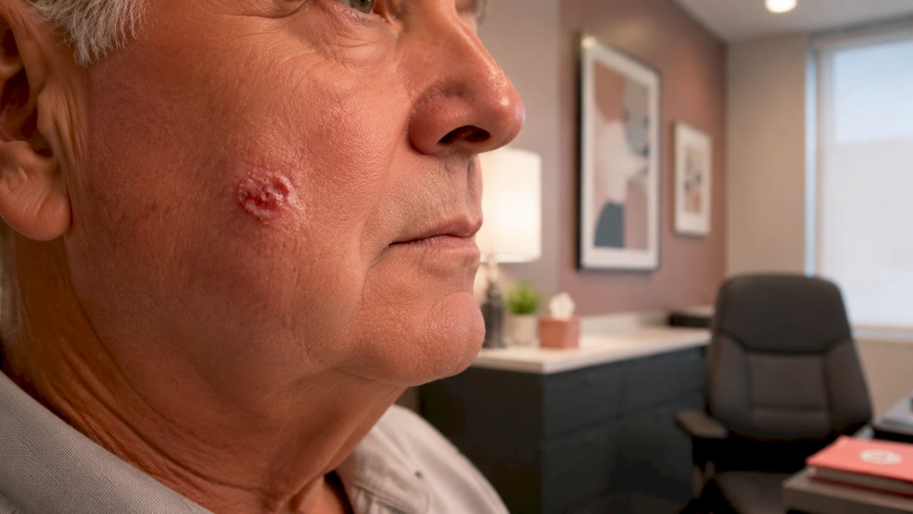
The three subtypes most relevant to delayed presentation are nodular, superficial, and infiltrative BCC. Nodular BCC tends to grow as a raised, pearly nodule that slowly enlarges and may develop a central ulcer. Superficial BCC spreads outward across the skin surface and can cover a wide area before it is noticed. Infiltrative BCC is the most clinically concerning subtype because it sends microscopic tendrils into the surrounding tissue well beyond the visible border of the lesion, making it far harder to remove completely.
Over a two-year period without treatment, the following changes are commonly observed:
- Increase in lesion diameter, often reaching 10–20 mm or larger depending on subtype and location
- Ulceration, where the centre of the lesion breaks down, bleeds intermittently, and fails to heal
- Crusting and scabbing that recurs repeatedly, often mistaken for a minor wound
- Colour changes, including areas of translucency, grey, or brown pigmentation within the lesion
- Border irregularity, with the edge becoming less defined as infiltrative growth advances
- Bleeding on contact, particularly when washing or drying the face
The location of the lesion matters considerably. BCCs on the nose, inner corner of the eye, ear canal, or scalp are classified as high-risk sites by the British Association of Dermatologists because the anatomy in these areas allows deeper invasion to occur with fewer visible warning signs.
Pro Tip: If a skin lesion has bled more than twice without an obvious cause, or has failed to heal over four weeks, seek a specialist assessment rather than waiting for a scheduled GP appointment. Early review changes outcomes.
What are the complications of leaving BCC untreated?
The physical complications of untreated BCC over two years extend well beyond the skin surface. Large tumours frequently exceed 30 mm and can invade muscle, cartilage, and bone, particularly when located on the face. Bone involvement, known as periosteal or cortical invasion, requires surgical approaches that include bone resection alongside skin reconstruction. This is a fundamentally different operation from the straightforward excision that would have been appropriate at an earlier stage.
Functional impairment is a real risk when BCC is located near critical anatomical structures. A tumour near the lower eyelid can distort the lid margin, impairing tear drainage and vision. A lesion on the nasal ala can erode the cartilage framework, affecting both breathing and appearance. BCCs adjacent to the ear canal have been documented to invade the external auditory meatus, causing conductive hearing loss.
The psychological burden of a visible, ulcerating lesion is also significant. Anxiety, social avoidance, and lowered self-esteem are well-documented consequences of untreated BCC, and these psychological effects often begin before the medical complications become severe. Patients frequently describe withdrawing from social situations, avoiding mirrors, and delaying treatment further because they feel embarrassed or frightened. This cycle of avoidance compounds the physical harm.
“The psychological distress caused by a visible BCC lesion can impair quality of life well before surgical intervention is considered. Holistic patient care must address both the physical and emotional dimensions of a delayed diagnosis.”
Advanced BCC cases most commonly present in older patients, and delayed presentation often involves tumours larger than 30 mm in anatomically complex facial regions such as the nose, orbit, and forehead. These are precisely the areas where reconstruction is most technically demanding and where the consequences of incomplete removal are most visible.
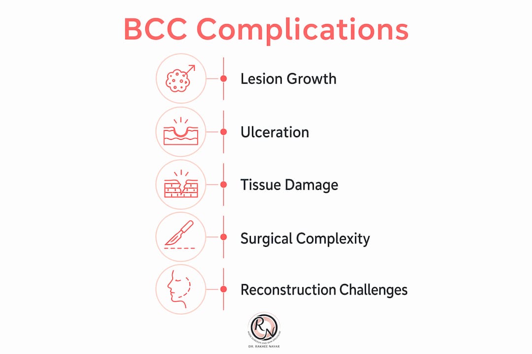
The key complications to be aware of include:
- Deep tissue invasion into subcutaneous fat, muscle, and periosteum
- Bone involvement, requiring partial resection of facial bones in severe cases
- Disfigurement, including loss of structural landmarks such as the nasal tip or eyelid margin
- Functional impairment, affecting vision, breathing, or hearing depending on tumour location
- Psychological distress, including anxiety and social withdrawal linked to visible lesion changes
- Increased recurrence risk, particularly with infiltrative subtypes that are difficult to clear completely
How does a two-year delay affect surgical options?
A two-year delay does not simply mean a larger wound. It changes the entire surgical equation. Delayed treatment significantly increases the likelihood that standard excision will be insufficient, and that complex reconstructive surgery will be required to restore both function and appearance.
Standard surgical excision with a 4–5 mm margin is appropriate for small, well-defined, low-risk BCCs. After two years of growth, the same lesion may now require margins of 10 mm or more, and the infiltrative subtype may have spread beyond any clinically visible border. In these circumstances, Mohs micrographic surgery offers a clear advantage. Mohs surgery removes the tumour layer by layer, examining 100% of the surgical margin under the microscope in real time. This approach reduces recurrence and preserves the maximum amount of healthy tissue, which is particularly valuable on the face.
The table below summarises the key differences between standard excision and Mohs surgery for delayed BCC cases:
| Factor | Standard Excision | Mohs Micrographic Surgery |
|---|---|---|
| Margin assessment | Sample of margin only | 100% of margin examined |
| Tissue preservation | Wider margins taken as standard | Maximum healthy tissue preserved |
| Suitability for delayed cases | Limited in infiltrative or large tumours | Preferred for complex, recurrent, or large BCCs |
| Reconstruction required | Often needed in delayed cases | Planned immediately after clearance |
| Recurrence rate (primary BCC) | Around 4–5% at five years | Lower for high-risk and recurrent cases |
| Cosmetic outcome | Variable | Optimised through tissue-sparing technique |
When a tumour has invaded the periosteum or bone, reconstruction becomes considerably more complex. Surgical treatment after two years frequently requires full-thickness skin grafts or local rotation flaps to close the defect. In cases involving bone resection, titanium mesh or cartilage grafts may be needed to restore structural support. Recovery time is longer, the risk of complications is higher, and the aesthetic outcome is harder to predict.
Pro Tip: Ask your surgeon specifically whether Mohs surgery is available for your case. For BCCs on the face, ears, or scalp, Mohs versus standard excision is a genuine clinical choice with measurable differences in outcome.
What should you do if your BCC has been untreated?
The single most important step is to seek a specialist consultation without further delay. Early specialist assessment prevents further tissue damage and improves both the surgical and aesthetic outcome. Waiting an additional month or two while considering options is unlikely to be catastrophic, but every additional period of growth adds to the complexity of treatment.
When you attend a specialist consultation, the clinician will assess the following:
- Lesion size and borders, including whether the edges are well-defined or irregular
- Subtype, confirmed by biopsy if not already done, as this determines the surgical approach
- Depth of invasion, assessed clinically and sometimes with imaging such as ultrasound or MRI for larger lesions
- Location risk, with high-risk sites on the face requiring more detailed surgical planning
- Reconstruction requirements, discussed before surgery so you understand the likely outcome
Alongside seeking treatment, there are practical steps you can take to support your skin health. Reducing UV exposure lowers the risk of new BCCs developing, and this applies whether or not you have had treatment. Wearing SPF 50 sunscreen daily, covering exposed skin, and avoiding peak sun hours between 11 am and 3 pm are all evidence-based measures. Regular skin checks, either self-examination monthly or with a specialist annually, allow new lesions to be identified at an earlier and more treatable stage.
Patient education about the signs of BCC progression reduces further delays in seeking care. Knowing what to look for, including a lesion that bleeds, crusts, grows, or fails to heal, means you are less likely to dismiss a new change as insignificant. You can review BCC warning signs visually to familiarise yourself with what different subtypes look like at various stages.
Key takeaways
Basal cell carcinoma left untreated for two years causes measurable tissue damage, increases surgical complexity, and carries significant psychological consequences that begin well before the physical complications become severe.
| Point | Details |
|---|---|
| BCC grows slowly but persistently | Typical growth of 2–4 mm per year means a two-year delay adds meaningful size and depth. |
| Deep invasion is a real risk | Untreated BCC can reach muscle, cartilage, and bone, particularly on the face. |
| Surgery becomes more complex | Two-year delays frequently require flap reconstruction or bone resection rather than simple excision. |
| Mohs surgery is preferred for delayed cases | It examines 100% of the surgical margin and preserves maximum healthy tissue. |
| Psychological impact begins early | Anxiety and social avoidance are documented consequences of visible, untreated lesions. |
Why i treat late-presenting BCC differently
In my clinical practice, the patients who present after two or more years without treatment are often the ones who understood their diagnosis but felt paralysed by it. They knew something was wrong. They had been told. But the combination of fear, uncertainty about what surgery would involve, and the slow-growing nature of BCC created a window in which inaction felt tolerable.
What I have observed consistently is that the surgical challenge in these cases is rarely the tumour itself. The challenge is the reconstruction. Removing a BCC that has grown for two years on the nose or lower eyelid is technically straightforward with Mohs surgery. Restoring the anatomy afterwards, in a way that preserves function and looks natural, is where the real complexity lies. This is why dual training in both Mohs surgery and plastic surgery matters so much for late-presenting cases. The excision and the reconstruction are not two separate problems. They are one operation, planned together from the outset.
The other thing I would say to anyone reading this is that the psychological weight of a visible, untreated lesion is underestimated in most clinical consultations. Patients arrive having avoided mirrors, cancelled social plans, and spent months convincing themselves the lesion is not as bad as it looks. Addressing that honestly, and explaining clearly what treatment will achieve, is as important as the surgery itself. A well-reconstructed face after Mohs surgery looks far better than the lesion it replaced. That is not a promise of perfection. It is a clinical reality I see in my patients regularly.
If you have a BCC that has been present for two years or more, the conversation with a specialist is not one to dread. It is the point at which the situation becomes manageable.
— Miss Rakhee Nayar
Expert BCC care at rakhee nayar – mohs surgeon and skin specialist
If you have a basal cell carcinoma that has been present for two years or longer, specialist assessment is the right next step.

Rakhee Nayar – Mohs Surgeon and Skin Specialist offers private, consultant-led care at Circle Cheshire in North West England, with e-consultations available for UK and international patients. Miss Nayar holds dual training in Mohs micrographic surgery and plastic surgery, meaning excision and reconstruction are planned together for the best possible outcome. Whether your BCC requires Mohs surgery or complex facial reconstruction, your care is directed by a GMC-registered Consultant Plastic Surgeon from first consultation to final review. To understand your options, begin with our guide to early detection benefits or book a consultation directly through the website.
This article is for informational purposes only and does not constitute medical advice. Please consult a GMC-registered specialist for assessment and treatment of any skin lesion.
FAQ
What happens if BCC is left untreated for 2 years?
Basal cell carcinoma left untreated for two years typically grows by 4–8 mm in diameter and may begin to ulcerate, bleed, and invade deeper tissue layers including muscle and bone. The longer it is left, the more complex the surgery required to remove it.
Can untreated BCC spread to other organs?
BCC very rarely metastasises to other organs, but it causes serious local destruction by invading surrounding skin, cartilage, and bone. The primary risk of untreated BCC is local tissue damage and disfigurement, not distant spread.
Is mohs surgery necessary for a BCC that has been untreated for 2 years?
Mohs micrographic surgery is the preferred approach for BCCs that are large, located on the face, or of an infiltrative subtype, all of which are more likely after a two-year delay. It offers the highest cure rates and preserves the most healthy tissue for reconstruction.
What are the risk factors for developing further bccs after treatment?
The main risk factors for skin cancer recurrence include cumulative UV exposure, fair skin, a history of previous BCCs, and immunosuppression. Ongoing sun protection with SPF 50 and annual skin checks significantly reduce the risk of new lesions.
Should i treat basal cell carcinoma even if it seems small?
Yes. Even a small BCC will continue to grow and may develop into a more complex lesion over time. Early treatment results in a simpler operation, a smaller scar, and a better cosmetic outcome than treatment delayed by months or years.