Skin cancer on the ear is a distinct clinical condition defined by the abnormal growth of malignant cells on the external ear, including the pinna, helix, lobule, and surrounding skin. The ear is one of the most commonly affected sites on the head and neck, largely because it receives continuous ultraviolet radiation with almost no natural shielding. The three main types found here are basal cell carcinoma (BCC), squamous cell carcinoma (SCC), and melanoma, each with different behaviour and treatment requirements. Recognising ear skin cancer signs early is the single most important factor in achieving a good outcome. Mohs micrographic surgery is the preferred treatment for most ear skin cancers, offering precise removal while preserving the delicate structures of the ear.
What are the symptoms of skin cancer on the ear?
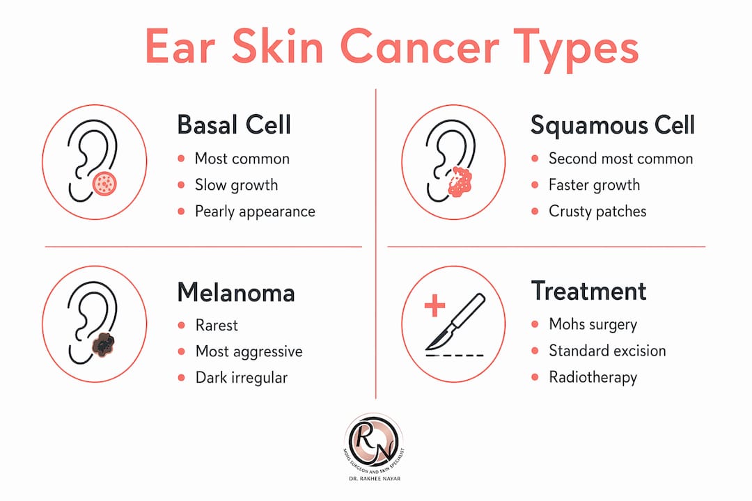
The ear presents three distinct types of skin cancer, each with recognisable features that set it apart from benign lesions.
Squamous cell carcinoma is the most common skin cancer found on the ear. It accounts for 0.2% of all face and neck cancers, which understates its significance given how frequently it appears specifically on the ear’s sun-exposed surfaces. SCC on the ear carries a higher risk of spreading to nearby lymph nodes than SCC elsewhere on the face. That makes prompt diagnosis and treatment non-negotiable.
Basal cell carcinoma is the most common skin cancer overall and the second most frequent type on the ear. It typically presents as a pearly or translucent lump, or a crusty spot that refuses to heal. BCC is frequently mistaken for a benign skin tag or dry patch, which delays diagnosis. It rarely spreads but can cause significant local destruction if left untreated on the ear’s cartilage.
Ear melanoma is rarer but the most aggressive of the three. It often appears as a new or changing dark patch, an irregular mole, or a lesion with uneven borders and variable colour. Melanoma on the ear demands urgent assessment because of its potential to spread rapidly to distant organs.
Common ear skin cancer signs to look for
The following symptoms warrant a professional assessment without delay:
- A sore or ulcer on the ear that has not healed within four weeks
- A scaly, crusty, or rough patch on the helix or lobule
- A firm, flesh-coloured, or pearly lump on the outer ear
- A dark or multicoloured patch that is new or has changed in size, shape, or colour
- Persistent bleeding or oozing from a spot on the ear
- Lumps near the ear or in the neck, which may indicate lymph node involvement
If an ear skin cancer is left untreated, advanced cases can affect the ear canal, cause hearing changes, or involve facial nerves. These complications are avoidable with early intervention.
How ear skin cancer types compare
| Feature | Basal Cell Carcinoma | Squamous Cell Carcinoma | Melanoma |
|---|---|---|---|
| Frequency on ear | Second most common | Most common | Rare |
| Typical appearance | Pearly lump, non-healing crust | Scaly, red, firm nodule | Dark, irregular patch |
| Risk of spread | Very low | Moderate to high | High |
| Urgency | High | Very high | Urgent |
Why is the ear particularly vulnerable to skin cancer?
The ear’s anatomy creates a near-perfect environment for UV damage to accumulate undetected. The skin over the cartilage is thin, tightly adherent, and offers little padding between the surface and the underlying structure. This means even a small tumour can reach cartilage quickly, complicating surgical removal.
The ear also sits in a position that receives direct and reflected UV radiation throughout the day. Unlike the nose or forehead, it lacks brow ridges or other anatomical features that provide partial shade. People who spend time outdoors, drive regularly, or work near windows accumulate significant UV exposure on the ear over decades, often without realising it.
Key risk factors for ear skin cancer
The following factors increase the likelihood of developing skin cancer on the ear:
- Fair skin, light eyes, or a history of sunburn: These individuals have less melanin to absorb UV radiation.
- Cumulative sun exposure: Decades of outdoor work or leisure activity without ear protection.
- Indoor tanning: Indoor tanning before age 35 increases melanoma risk by 75%. Even a single session raises it by 20%.
- Previous skin cancers: A history of BCC or SCC elsewhere on the body significantly raises the risk of a new lesion on the ear.
- Immunosuppression: Organ transplant recipients and those on long-term immunosuppressant therapy face substantially elevated risk.
- Chronic ear conditions: Longstanding inflammation or scarring of the ear skin can predispose to SCC in particular.
- Male sex: Men are statistically more likely to develop ear skin cancer, partly due to shorter hair offering less coverage.
Clouds do not eliminate UV risk. Up to 80% of harmful UV rays can penetrate cloud cover, meaning overcast days still require sun protection. This is a fact many patients find surprising and which directly explains why ear cancers develop even in people who avoid obvious sun exposure.
Pro Tip: When applying sunscreen to the face, most people miss the helix (the outer rim of the ear), the back of the ear, and the area just behind the earlobe. Apply sunscreen to these areas specifically, using a fingertip to work it into the folds.
How is ear skin cancer diagnosed and treated?
Diagnosis: from self-examination to biopsy
Diagnosis begins with a thorough clinical examination by a GMC-registered specialist. Annual skin checks are the standard recommendation for anyone with a history of skin cancer or significant UV exposure. During an assessment, the clinician examines the entire ear, including the posterior surface and the skin behind the ear, areas that patients and even some clinicians overlook.
A biopsy remains the definitive diagnostic step for most lesions. A small sample of tissue is taken under local anaesthetic and sent for histological analysis. In some cases, teledermatology can diagnose BCC and SCC from high-quality digital images, enabling direct treatment without a preliminary biopsy. This approach reduces the number of procedures a patient needs and speeds up the pathway to treatment.
Surgical treatment options for ear skin cancer
Three main surgical approaches are used for ear skin cancers:
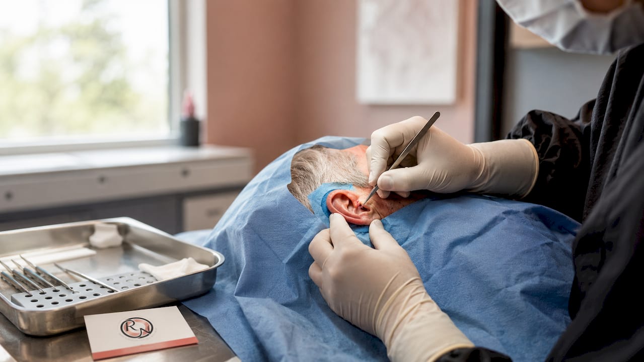
- Mohs micrographic surgery: The gold standard for most ear skin cancers. The surgeon removes the tumour layer by layer, examining each layer under a microscope before proceeding. This continues until all margins are clear. The result is complete tumour removal with the smallest possible wound.
- Standard surgical excision: The tumour is removed with a margin of surrounding tissue. Margins are checked after the procedure, which means a second operation is sometimes needed if the margins are not clear.
- Wedge excision: A triangular section of the ear is removed to include the tumour and a clear margin. Most wedge excisions are performed under local anaesthetic, avoiding the risks associated with general anaesthesia. This is particularly relevant for older patients or those with significant health conditions.
When surgery is not suitable, radiotherapy offers an alternative. It is used for patients who cannot tolerate surgery or where the tumour location makes complete surgical removal difficult. Topical treatments such as imiquimod or fluorouracil are occasionally used for very superficial BCC but are not appropriate for invasive cancers on the ear.
Mohs surgery versus standard excision: a direct comparison
Clinical guidelines recommend Mohs surgery for ear skin cancers specifically because the ear’s anatomy demands tissue preservation alongside complete tumour removal. The table below summarises the key differences.
| Factor | Mohs Micrographic Surgery | Standard Excision |
|---|---|---|
| Margin assessment | Real-time, 100% of margin examined | Delayed, representative sampling only |
| Tissue preservation | Maximum | Standard margins removed |
| Cure rate (primary BCC) | Up to 99% | Up to 95% |
| Risk of second operation | Very low | Higher if margins involved |
| Suitability for ear | Preferred | Suitable for low-risk lesions |
| Anaesthetic | Local | Local or general |
For a detailed breakdown of how these two approaches differ in practice, the Mohs vs standard excision guide on the Rakhee Nayar – Mohs Surgeon and Skin Specialist website covers the clinical decision points clearly.
Reconstruction after ear surgery
Removing a skin cancer from the ear often leaves a defect that requires careful reconstruction. The goal is to restore both the appearance and function of the ear. Options include direct closure, skin grafts, and local flaps, depending on the size and location of the defect. Miss Nayar’s dual training in Mohs surgery and plastic surgery means that reconstruction is planned from the outset, not as an afterthought. Patients considering their cosmetic outcomes after surgery will find that Mohs surgery consistently produces smaller defects and better aesthetic results than standard excision.
Pro Tip: Ask your surgeon at the initial consultation whether reconstruction will be performed on the same day as tumour removal. For ear lesions, same-day reconstruction is usually possible with Mohs surgery and reduces the total number of procedures.
How can you reduce your risk of ear skin cancer?
Prevention is a year-round commitment, not a seasonal one. The following steps directly reduce UV damage to the ear and lower the risk of skin cancer developing or recurring.
- Apply broad-spectrum sunscreen daily. Use SPF 30 or higher on all exposed skin, including the ears. SPF 30 blocks approximately 97% of UVB rays when applied correctly and reapplied every two hours. Most people apply far too little to achieve the labelled protection.
- Wear a wide-brimmed hat. Wide-brimmed hats offer superior coverage for the ears compared to baseball caps, which leave the ears fully exposed. Choose hats with a brim of at least 7.5 centimetres made from tightly woven fabric.
- Avoid peak UV hours. UV radiation is strongest between 10 a.m. and 4 p.m. Seek shade during these hours when outdoors for extended periods.
- Do not use tanning beds. Indoor tanning significantly raises melanoma risk, as noted above. There is no safe level of UV exposure from a tanning bed.
- Examine your ears regularly. Use a hand mirror and a well-lit room to check the outer rim, the bowl of the ear, the back of the ear, and the skin behind the earlobe. Look for any new or changing lesion. If you notice anything unusual, seek a professional assessment promptly.
- Attend annual skin checks. Anyone with a personal or family history of skin cancer, significant sun exposure, or fair skin should see a specialist annually. A trained clinician will examine areas you cannot easily assess yourself.
For broader guidance on skin cancer prevention tips and recovery, the Rakhee Nayar – Mohs Surgeon and Skin Specialist website provides detailed, clinically grounded resources.
Key takeaways
Skin cancer on the ear requires early recognition, specialist assessment, and, in most cases, Mohs micrographic surgery to achieve the highest cure rates while preserving the ear’s structure and appearance.
| Point | Details |
|---|---|
| SCC is the most common ear cancer | Squamous cell carcinoma on the ear carries a higher risk of spread than SCC elsewhere on the face. |
| Mohs surgery is the preferred treatment | It examines 100% of the surgical margin in real time, minimising recurrence and tissue loss. |
| The ear is a high-risk UV site | Thin skin, no natural shading, and frequent neglect during sunscreen application make the ear vulnerable. |
| Prevention requires year-round effort | SPF 30 sunscreen, wide-brimmed hats, and avoidance of tanning beds reduce risk significantly. |
| Early detection changes outcomes | Annual skin checks and prompt assessment of non-healing lesions are the most effective protective steps. |
What i have learnt treating ear skin cancers
The ear is the site I find patients most consistently underestimate. They notice a crusty spot on the helix, assume it is dry skin or an old scar, and wait. By the time they attend a consultation, weeks or months have passed. In a few cases, that delay has meant the tumour has reached cartilage, which changes the reconstruction entirely.
What I want patients to understand is that ear skin cancers are almost always treatable when caught early. The anatomy is complex, but that complexity is precisely why Mohs surgery was developed and why it performs so well here. Removing a tumour layer by layer, with real-time microscopic margin control, means I can clear the cancer completely while leaving as much healthy tissue as possible. For an ear, where every millimetre of skin matters for both appearance and structure, that precision is not a luxury. It is the standard of care.
I also want to address a concern I hear frequently: patients worry that ear surgery will leave them with a visible deformity. In the majority of cases, with careful planning and same-day reconstruction, the cosmetic result is far better than patients anticipate. The cure rates achieved with Mohs surgery are high, and the aesthetic outcomes, when reconstruction is integrated from the outset, are genuinely good.
My practical advice is this: if you have a spot on your ear that has not healed in four weeks, do not wait for your next routine appointment. Book a specialist assessment. The earlier the diagnosis, the simpler the treatment, and the better the result.
This article is for informational purposes only and does not constitute medical advice. Please consult a GMC-registered specialist for assessment and treatment of any suspicious lesion.
— Miss Rakhee Nayar
Expert mohs surgery for ear skin cancer at rakhee nayar – mohs surgeon and skin specialist
If you have noticed a suspicious lesion on your ear, or have already received a diagnosis and want to understand your treatment options, Rakhee Nayar – Mohs Surgeon and Skin Specialist offers consultant-led assessment and treatment at Circle Cheshire in North West England.

Miss Nayar is a GMC-registered Consultant Plastic Surgeon, FRCS (Plast), with dual training in Mohs micrographic surgery and plastic surgery. She provides private consultations, e-consultations for patients across the UK and internationally, and same-day Mohs surgery with reconstruction for eligible patients. Begin with the skin cancer detection guide to understand what to expect, or visit the Mohs surgery service page to learn how treatment is delivered. For a full overview of what to look for, the UK skin cancer symptoms guide is a thorough starting point.
FAQ
What does skin cancer on the ear look like?
Skin cancer on the ear most commonly appears as a non-healing sore, a crusty or scaly patch, a pearly lump, or a dark irregular lesion on the outer ear. Any spot that has not resolved within four weeks should be assessed by a specialist.
Is mohs surgery the best treatment for ear skin cancer?
Mohs micrographic surgery is the recommended treatment for most ear skin cancers because it removes the tumour with real-time margin control, preserving as much healthy tissue as possible. This is particularly important on the ear, where tissue loss affects both appearance and structure.
Can ear skin cancer spread to other parts of the body?
Squamous cell carcinoma on the ear carries a higher risk of spreading to nearby lymph nodes than SCC on other facial sites. Melanoma can spread to distant organs. Basal cell carcinoma rarely spreads but can cause significant local destruction if untreated.
How do i check my own ears for skin cancer signs?
Use a hand mirror in good light to examine the outer rim, the bowl, the back of the ear, and the skin behind the earlobe. Look for any new lesion, a spot that has changed, or any sore that has not healed. Ask a partner or family member to check areas you cannot see clearly.
What is the cost of private mohs surgery for ear skin cancer in the UK?
Private Mohs surgery costs vary depending on the size and complexity of the tumour and the reconstruction required. Consultation fees and surgical costs should be discussed directly with the clinic. Rakhee Nayar – Mohs Surgeon and Skin Specialist provides transparent pricing information at the time of consultation.