TL;DR:
- Persistent facial skin lesions should be evaluated promptly for early detection of basal cell carcinoma.
- Early treatment options, especially Mohs surgery, offer high cure rates with minimal scarring.
- Regular monitoring and professional assessment are crucial for suspicious spots that do not heal or change over time.
Noticing a mark on your face that refuses to heal can be unsettling. You might dismiss it as a slow-healing spot or a patch of dry skin, but persistent facial lesions deserve closer attention. Basal cell carcinoma is the most common facial skin cancer, appearing as a pink or pearly bump, scab, or ulcer on sun-exposed areas. The good news is that when identified early, treatment is highly effective and cosmetic outcomes are far better. This guide walks you through recognising the warning signs, taking the right next steps, and understanding what treatment involves so you can act with confidence rather than anxiety.
Key Takeaways
| Point | Details |
|---|---|
| BCC is common on the face | Regularly check your face for lasting or changing marks, especially if you have fair skin or a history of sun exposure. |
| Spot warning signs early | Look for non-healing sores, pearly bumps, or scaly patches and consult your GP promptly. |
| Early treatment protects appearance | Prompt diagnosis and specialist care, such as Mohs surgery, offer the best cosmetic results and cure rates. |
| Support is available | Expert help and advanced facial treatments are accessible in the UK for those at risk or newly diagnosed. |
What is basal cell carcinoma and why is facial identification vital?
Basal cell carcinoma is a type of non-melanoma skin cancer that develops in the basal cells, the deepest layer of the epidermis. It grows slowly, rarely spreads to other parts of the body, but left untreated it can burrow into surrounding tissue, nerves, and even bone. On the face, that destruction can be significant and distressing.
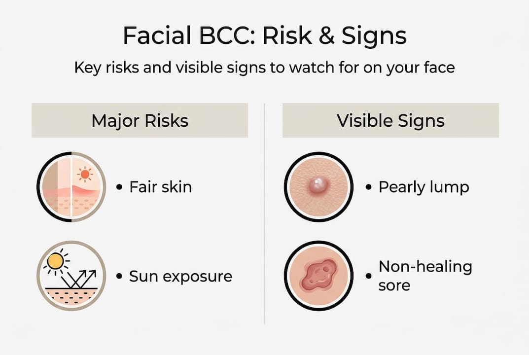
The primary cause is cumulative ultraviolet radiation. Every summer holiday, every unprotected lunch break, every decade of British sun adds up. The face is most exposed, which is why 3 in 10 fair-skinned UK adults develop BCC in their lifetime, with the face as the most common site.
People with skin type I or II (pale skin that burns easily), those over 50, and anyone with a history of significant sun exposure or sunbed use carry the highest risk. That said, BCC is not exclusively an older person’s problem. Cases in people in their 30s and 40s are increasingly common.
Who is most at risk?
- Fair skin that burns rather than tans
- History of repeated sunburn or sunbed use
- Living or working outdoors for many years
- Previous personal or family history of skin cancer
- Immunosuppression due to medication or illness
| Risk factor | Level of impact |
|---|---|
| Fair skin (type I/II) | High |
| Cumulative UV exposure | High |
| Age over 50 | Moderate to high |
| Immunosuppression | Moderate |
| Family history of skin cancer | Moderate |
The crucial point is this: BCC caught early is overwhelmingly treatable. When a lesion has grown large or invaded deeper structures on the face, treatment becomes more complex, scarring increases, and reconstruction may be necessary. Knowing skin cancer symptoms on the face before a lesion has a chance to advance is the single most important step you can take for both your health and your appearance.
How to recognise basal cell carcinoma on your face: visible signs and symptoms
Not every BCC looks the same, and that is precisely what makes them tricky to spot. Understanding the main types is essential.
The three main types of facial BCC:
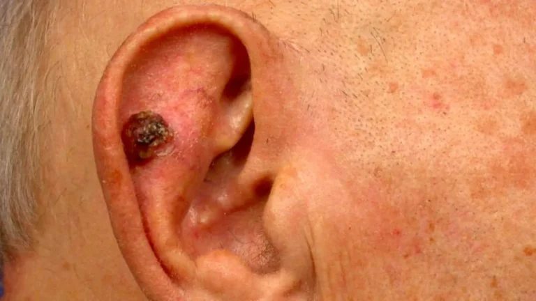
- Nodular BCC is the most common. It appears as a shiny, pearly, or translucent bump, often with tiny red blood vessels running across its surface. It may develop a central ulcer that bleeds and crusts, then appears to heal before breaking down again.
- Superficial BCC looks more like a scaly, pink or red flat patch. It can resemble eczema or a patch of irritated skin. It tends to spread outwards rather than downwards.
- Morphoeic (sclerosing) BCC is the most deceptive. It appears as a flat, pale, scar-like area with poorly defined edges. Many patients and even some GPs initially mistake it for a minor scar or stretch mark.
BCC can appear as a pink or red pearly lump, a sore that will not heal, a scaly patch, or an ulcer, often with tiny red blood vessels visible at the surface.
What to look for when checking your face:
- A spot or sore that has not healed after four weeks
- A shiny or pearlescent bump, even if small
- A pink or red patch with a slightly raised or rolled edge
- A lesion that bleeds, crusts, and appears to partially heal repeatedly
- A pale, flat, scar-like area you cannot explain
- Visible tiny red blood vessels (telangiectasia) across a lesion
Important: Morphoeic BCC is frequently underestimated because it looks so innocuous. If you have a pale, flat mark on your face that has been there for months without a clear cause, do not ignore it.
Pro Tip: Take a well-lit, close-up photo of any suspicious area monthly. Comparing photos over six to eight weeks makes it much easier to spot subtle changes in size, colour, or texture that the eye alone might miss.
Regular skin cancer detection checks, especially of the face, nose, ears, and scalp, are worthwhile for anyone over 40 with a history of sun exposure. The ABCDE check used for melanoma can be adapted for BCC: focus on any non-healing, growing, or bleeding lesion as a prompt to act.
For more detail on spotting face lesions and understanding what normal versus abnormal looks like, specialist resources can help you build confidence in your own assessments. The Skin Health Info resource also provides useful visual references.
What to do if you spot a suspicious spot: next steps and diagnosis
Spotting something that concerns you is only half the task. Acting on it promptly is the other half.
Step-by-step guide once you notice a suspicious area:
- Photograph the area clearly in good lighting. Note the date.
- Continue photographing weekly to document any changes in size, bleeding, or appearance.
- Book an appointment with your GP. Do not wait to see if it resolves after eight weeks.
- At the appointment, show your photos. Describe how long it has been there and whether it has bled or changed.
- Ask your GP specifically whether a dermatology referral is warranted.
In the UK, see a GP promptly about any non-healing sore or changing facial lesion. NHS referral for suspected BCC typically falls within 18 weeks, though suspected high-risk cases may be fast-tracked on the two-week wait pathway.
Do not let appointment anxiety hold you back. GPs see skin lesions regularly and referral is a routine process. Bring your photographs, keep a brief written note of when you first noticed the lesion, and ask whether a skin biopsy will be needed.
What to expect after referral:
- Clinical examination by a dermatologist or specialist
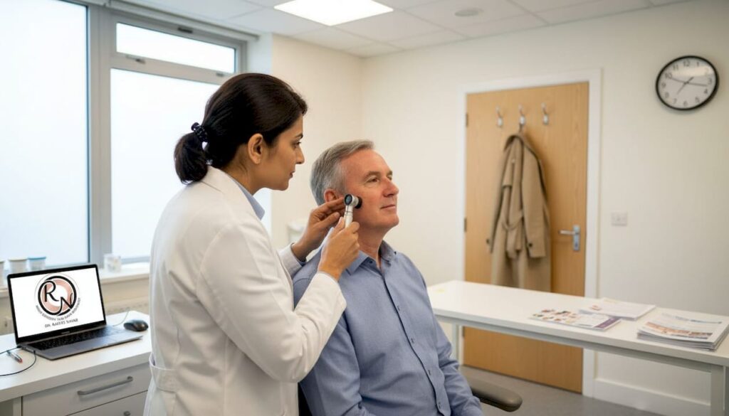
- Dermoscopy (magnified examination using a handheld lens)
- Skin biopsy performed under local anaesthetic to confirm diagnosis
Diagnosis is confirmed through clinical appearance and a biopsy, which is a quick outpatient procedure and not as daunting as it sounds.
Pro Tip: Prepare three questions before your GP or specialist appointment: When was this likely to have started? What type of BCC is this, if confirmed? And what are my treatment options given its location on my face?
Good records and clear communication speed up the pathway to detecting skin cancer accurately and getting the right treatment in place. Once a diagnosis is confirmed, your specialist will discuss follow-up after diagnosis and what to expect next.
Treatment options for facial basal cell carcinoma and what to expect
Once your diagnosis is confirmed, the treatment chosen will depend on the type, size, location, and whether the BCC is primary or has previously been treated.
Main treatment options:
- Surgical excision: The standard approach. The lesion is removed with a margin of healthy tissue and sent for laboratory analysis.
- Mohs micrographic surgery: The gold standard for facial BCCs, particularly near the eyes, nose, lips, and ears. Tissue is removed layer by layer and examined under a microscope in real time until clear margins are confirmed.
- Topical treatments: Creams such as imiquimod or fluorouracil are used for superficial BCCs in lower-risk areas.
- Photodynamic therapy (PDT): Light-activated treatment suitable for superficial, non-aggressive BCCs.
- Curettage and cautery: Scraping and burning technique, appropriate for small, low-risk lesions.
- Radiotherapy: An option for patients who cannot undergo surgery.
Surgical excision is the main treatment, with Mohs micrographic surgery recommended for high-risk facial BCC, achieving cure rates above 98%.
Early BCC is highly curable. Alternatives for superficial types include creams, photodynamic therapy, and radiotherapy, particularly where surgery is not suitable.
For facial BCC close to critical structures, Mohs surgery is especially valuable because it removes the minimum amount of tissue necessary while ensuring complete clearance. This matters enormously for reconstruction. The less healthy tissue removed, the more natural the cosmetic result.
Pro Tip: If your BCC is on or near your nose, eyelids, lips, or ears, ask specifically about Mohs surgery for BCC. Standard excision margins in these areas can significantly affect appearance and function.
Reconstruction after surgery is a consideration worth raising early. Surgeons with dual training in Mohs and plastic surgery can plan excision and reconstruction after surgery as a single coordinated process, achieving far better results than treating them separately. Guidance on how to minimise scarring after removal is also part of the conversation you should be having before any procedure.
A specialist’s perspective: what most people miss about spotting and treating facial BCC
The lesions we worry least about are often the ones that cause the most trouble. Morphoeic BCC is the perfect example. Patients describe it as “just a scar from years ago” or “a flat mark that’s always been there.” It looks nothing like a cancer. That is the problem.
Clinically, the biggest mistake we see is not the failure to spot something dramatic; it is the assumption that a subtle, unchanging mark is therefore harmless. BCC does not always bleed, ulcerate, or draw attention to itself. Some of the most invasive cases we treat began as a pale, flat patch that a patient monitored for two or three years before seeking advice.
The other misconception worth challenging is the fear that surgery will cause more scarring than leaving things alone. In reality, the reverse is true. Mohs surgery, by preserving healthy tissue precisely, typically produces a far smaller and better-positioned scar than a BCC that has been allowed to enlarge. Understanding the facts about Mohs surgery early helps patients make better, calmer decisions. Early referral, an honest conversation with your specialist, and trust in the process produce the best outcomes, both medically and cosmetically.
Expert support and advanced treatments for facial BCC
If you have noticed a suspicious mark on your face, or if you have already received a BCC diagnosis and want to understand your options more fully, specialist input can make a significant difference to both your outcome and your peace of mind.

At mohssurgeon.co.uk, Miss Rakhee Nayar combines specialist training in both Mohs surgery and plastic surgery to offer patients in the UK a level of precision and reconstructive expertise that is genuinely rare. Whether you need thorough skin cancer detection, dedicated Mohs surgery for BCC on a high-risk facial site, or advice about facial reconstruction options, private consultations and e-consultations are available. You do not have to wait and worry. Expert guidance is accessible now.
Frequently asked questions
What does basal cell carcinoma look like on the face?
BCC on the face commonly appears as a pink or pearly lump that does not heal, a scab that bleeds repeatedly, an ulcer, or a scaly flat mark, sometimes with tiny visible red blood vessels across the surface.
How serious is facial basal cell carcinoma?
BCC rarely metastasises in fewer than 0.1% of cases, but it can locally destroy facial tissue if left untreated. Early detection leads to highly effective and cosmetically favourable treatment.
How can I tell if my spot needs to be checked by a doctor?
If any facial area does not heal, bleeds, grows, or changes over several weeks, see your GP. The NHS advises prompt assessment for any non-healing sore or unexplained skin change on the face.
Will Mohs surgery leave a noticeable scar on my face?
Mohs surgery is specifically designed to preserve as much healthy tissue as possible, resulting in minimal, well-positioned scars. It is the preferred technique for high-risk facial BCC, with cure rates above 98%.
Recommended
- How to Spot Skin Cancer Symptoms on the Face – Expert Mohs Surgery | UK
- SCC: Diagnosis, Mohs surgery and facial reconstruction
- Skin Cancer Detection – Expert Mohs Surgery | UK
- Mohs Micrographic Surgery for Basal Cell Carcinoma – Expert Mohs Surgery | UK
Filed under Spotting Skin Cancer