TL;DR:
- A skin lesion is any abnormal skin area that looks or feels different from its surroundings. Recognizing warning signs and understanding lesion types enable early detection and proper medical evaluation, especially for suspicious changes. Acting promptly when a lesion exhibits ABCDE features improves treatment outcomes and long-term skin health.
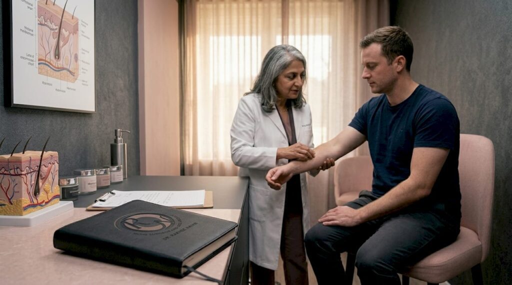
If you have ever noticed an unusual spot, bump, or patch on your skin and felt a wave of anxiety, you are not alone. A skin lesion is simply any area of skin that looks or feels different from the surrounding tissue. It can be raised or flat, coloured or pale, smooth or rough. The term covers everything from a harmless freckle to something that warrants medical attention. Understanding the difference is not about frightening yourself. It is about being informed enough to act calmly and correctly.
Key takeaways
| Point | Details |
|---|---|
| Broad medical term | A skin lesion describes any abnormal skin area, ranging from benign moles to potentially serious growths. |
| Many types exist | Lesions are classified by shape, size, elevation, and fluid content, each with distinct clinical significance. |
| Causes vary widely | UV exposure, infections, inflammation, trauma, and genetics all contribute to lesion formation. |
| ABCDE rule matters | Asymmetry, border, colour, diameter, and evolution are the key signs that distinguish benign from suspicious lesions. |
| Early evaluation saves lives | Seeking professional assessment promptly when warning signs appear significantly improves treatment outcomes. |
Types of skin lesions explained
Clinicians use a precise vocabulary to describe lesions, and understanding these terms helps you communicate accurately with a doctor. Medical morphology classifies lesions into categories based on size, elevation, and whether they contain fluid.
| Lesion type | Appearance | Size | Fluid present? |
|---|---|---|---|
| Macule | Flat colour change, no raised edge | Under 1 cm | No |
| Papule | Raised solid bump | Under 1 cm | No |
| Plaque | Flat-topped raised area | Over 1 cm | No |
| Nodule | Deep solid lump | Over 1 cm | No |
| Vesicle | Small blister | Under 1 cm | Yes (clear) |
| Pustule | Raised lesion with pus | Varies | Yes (cloudy) |
| Ulcer | Break in skin surface, often crusted | Varies | Sometimes |
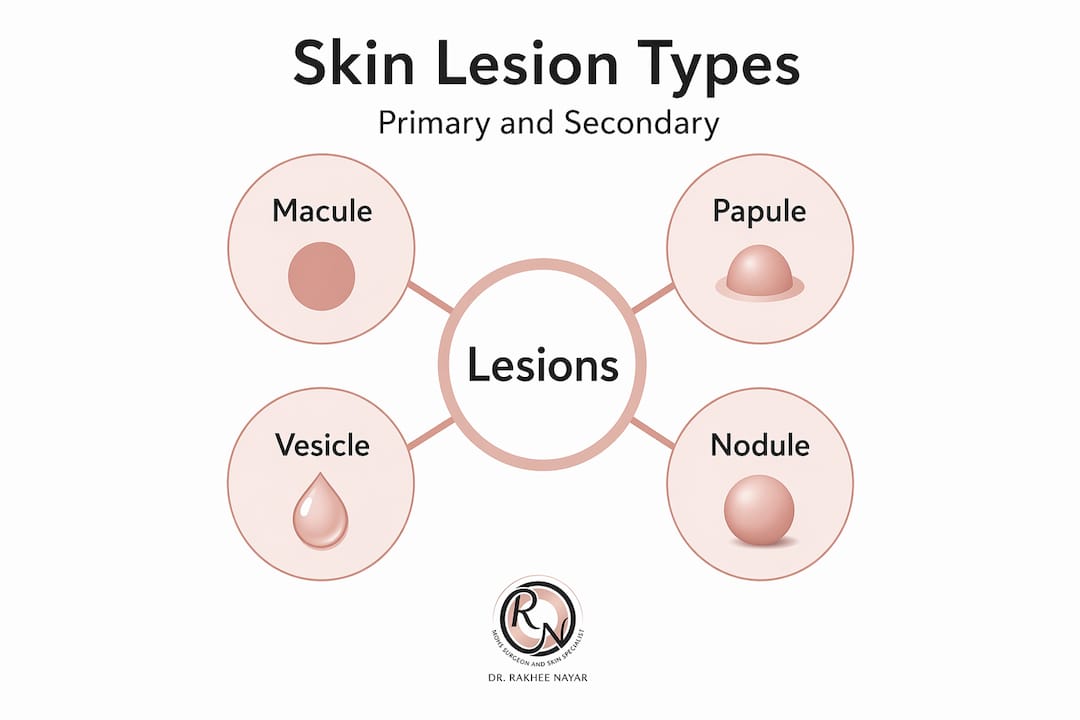
A macule is what you see with most freckles and flat birthmarks. A papule is what forms with acne spots or early warts. Plaques appear prominently in psoriasis, where raised patches of scaly skin develop over larger areas. Nodules are deeper and firmer, sometimes felt more than seen.
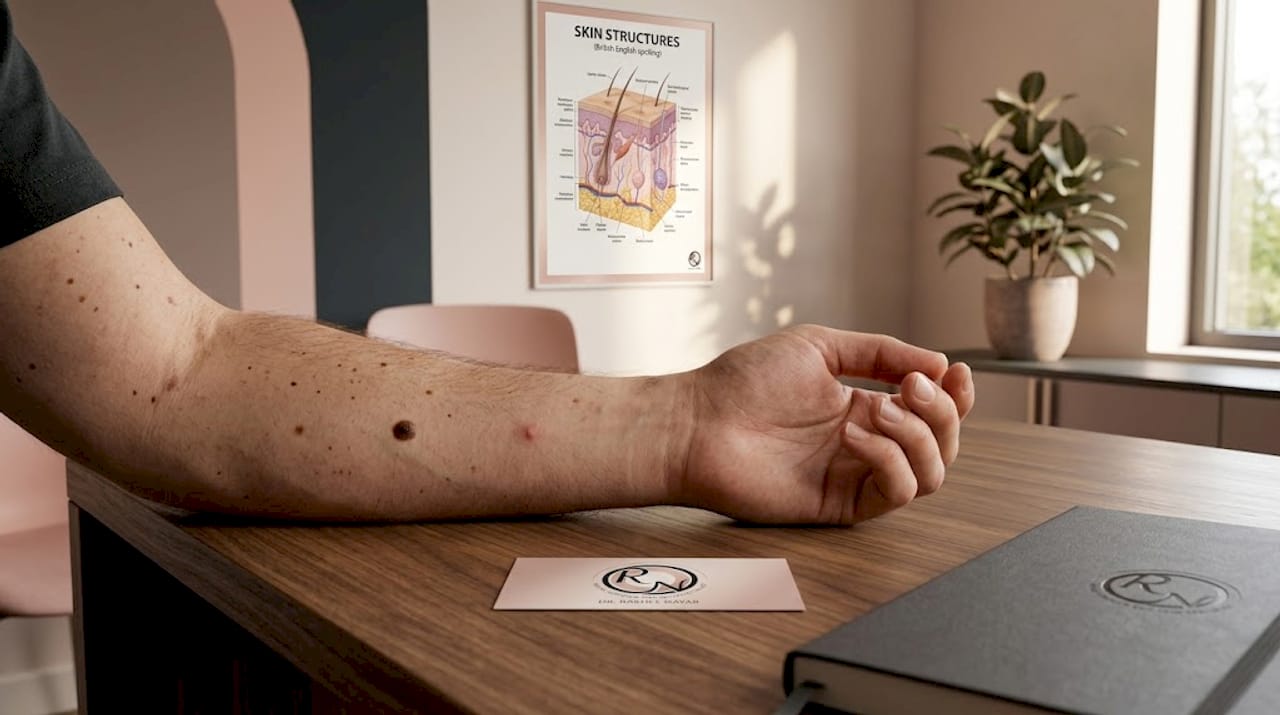
Beyond these clinical categories, you will encounter well-known examples in everyday life. Moles are pigmented macules or papules that most adults carry without concern. Seborrheic keratoses are common in middle-aged and older adults, appearing as waxy, stuck-on patches that look almost pasted onto the skin. Warts, caused by the human papillomavirus (HPV), are rough papules typically 2–10 mm in diameter that often resolve on their own over time.
Pro Tip: When examining a lesion yourself, use good natural lighting and run a fingertip gently across the surface. The distinction between flat and raised, smooth and rough, gives your doctor useful information before they even look at it.
What causes skin lesions
Understanding what causes skin lesions helps you recognise your own risk and take meaningful steps toward prevention. The origins are genuinely diverse.
- UV radiation. UV exposure damages skin DNA and is the primary driver behind many skin cancers, including basal cell carcinoma and squamous cell carcinoma. It also accelerates benign age spots and solar lentigines.
- Infections. Bacterial infections can cause impetigo (crusted sores) or folliculitis (infected hair follicles). Viral infections produce warts and cold sores. Fungal infections manifest as ring-shaped scaly patches like ringworm, while some parasites cause intensely itchy burrow lines seen in scabies.
- Inflammatory and autoimmune conditions. Eczema, psoriasis, and lupus all produce characteristic lesions as the immune system attacks skin cells or triggers chronic inflammation.
- Trauma and allergic reactions. A bruise is a lesion. So is a blister from a tight shoe or a raised hive following contact with an allergen. These are among the most common and self-limiting types you will encounter.
- Genetic predispositions. Some individuals are born with a higher density of melanocytes, producing more moles. Others carry gene variants that raise their lifetime risk of melanoma or other skin cancer types.
- Neoplastic processes. Both benign tumours (such as lipomas or dermatofibromas) and malignant ones arise from abnormal cell growth. These can be slow-growing and harmless or rapidly progressing and dangerous.
Pro Tip: Take stock of your personal risk profile. Fair skin, a family history of skin cancer, frequent sunburns in childhood, or a history of using sunbeds all raise your baseline risk. Knowing this helps you decide how vigilantly to monitor any new lesion.
Skin lesion symptoms and warning signs
Most lesions cause no symptoms at all. That is actually one reason why skin cancer is often caught late. Knowing when a lesion warrants concern depends on learning what normal looks like for your own skin and noticing when something changes.
The ABCDE framework is the most reliable starting point for identifying potentially malignant lesions, particularly melanoma. ABCDE stands for asymmetry (one half does not mirror the other), border irregularity (edges are notched or blurred), colour variation (multiple shades of brown, black, red, or white in one lesion), diameter greater than 6 mm, and evolution (any change in size, shape, or colour over time). The E is arguably the most telling. A stable mole you have had since childhood is almost always benign. One that is visibly different from a year ago deserves professional eyes.
Beyond the ABCDE checklist, watch for these specific warning signs:
- A sore or wound that does not heal within three to four weeks
- Bleeding from a lesion without any injury to cause it
- Persistent itching, tenderness, or pain within or around a lesion
- A firm, shiny, or pearly nodule (a hallmark of basal cell carcinoma)
- Rapid growth over days or weeks rather than months
- Crusting or scabbing that keeps returning after the area appears to heal
Dermoscopy allows clinicians to evaluate pigmented lesions on areas like the hands and feet in far more detail than the naked eye allows, reducing the number of unnecessary biopsies while catching genuine threats earlier. If you have a lesion in an acral site (palm, sole, or under a nail) that appears pigmented, this is precisely the kind of assessment you should ask about.
Seek medical evaluation without delay if any lesion meets the ABCDE criteria, shows the symptoms above, or simply feels different from anything you have noticed before. When it comes to skin changes, earlier is always better. Early detection changes outcomes in a measurable and often decisive way.
Diagnosis and treatment options
When you see a clinician about a lesion, the assessment follows a logical process. Visual examination comes first, often augmented by dermoscopy. Clinicians prioritise lesion morphology alongside your personal risk factors to decide whether monitoring, biopsy, or immediate treatment is appropriate.
Understanding the distinction between primary and secondary lesions is useful here. Primary lesions arise on previously normal skin. Secondary lesions develop from changes to primary ones, such as a vesicle that ruptures and becomes a crusted ulcer. This distinction shapes diagnosis because a secondary lesion tells a clinician what the skin has already been through.
When biopsy is indicated, a small sample of tissue is removed under local anaesthetic and examined under a microscope. This histopathological confirmation is the definitive step in diagnosing skin cancer. The type of biopsy (shave, punch, or excisional) depends on the lesion’s size, depth, and location.
| Treatment type | Best suited for | Key benefit |
|---|---|---|
| Observation and monitoring | Benign, stable lesions | Avoids unnecessary procedures |
| Cryotherapy | Small benign lesions (warts, keratoses) | Fast, minimal scarring |
| Standard surgical excision | Most skin cancers | Effective removal with margin |
| Mohs micrographic surgery | Facial/cosmetically sensitive skin cancers | Highest cure rate, tissue-sparing |
| Radiotherapy | Inoperable or high-risk lesions | Non-surgical option |
| Topical treatments | Superficial basal cell carcinoma | No surgery required |
Skin cancer treatments range from topical creams for superficial lesions to advanced surgical techniques for complex cases. Mohs micrographic surgery is particularly significant for lesions on the face, ears, and nose, where preserving healthy tissue matters enormously. After removal of a malignant lesion, reconstructive surgery may be recommended to restore both function and appearance.
Benign lesions do not always need removal. A seborrheic keratosis, for example, requires no treatment unless it becomes irritated or cosmetically bothersome. This is worth knowing because the decision to treat is not always about cancer risk. It can simply be about your quality of life.
My perspective on skin lesion anxiety
I have seen the pattern play out many times. Someone discovers a spot on their arm that has been there for years, suddenly notices it, and spends two weeks in quiet dread before doing anything about it. Then they see a doctor, who tells them it is a harmless seborrheic keratosis. The anxiety was real. The lesion was not dangerous. But those two weeks were wasted.
What I have learned is that knowledge is genuinely protective, not in a clichéd way but in a practical one. People who understand what a skin lesion actually is tend to react more proportionately. They know the difference between a stable mole and a rapidly changing nodule. They know the ABCDE rule. They know when to book an appointment versus when to simply observe.
The mistake I see most often is not panic. It is dismissal. People minimise lesions they should have checked earlier because they assume anything benign-looking must be fine. The problem is that many early-stage skin cancers look unremarkable. A pale, slightly shiny patch on the nose does not scream danger. But in my experience, those are precisely the cases where skin cancer detection makes the most difference.
My take is this: you do not need to be anxious about every mark on your skin. But you do need a baseline understanding of what you are looking at, and you need to act when something genuinely changes. Calm vigilance, not fear, is what keeps skin healthy long-term.
— Gregg
When to consider specialist skin care
If a lesion is concerning you, or if a GP has already flagged something for further review, specialist evaluation can provide clarity and real peace of mind.

Mohssurgeon offers expert skin cancer diagnosis and treatment through Miss Rakhee Nayar, who holds dual training in both plastic surgery and Mohs micrographic surgery. For lesions on the face or other sensitive areas, Mohs surgery provides the highest available cure rate while preserving as much healthy tissue as possible. Where reconstruction is needed following removal, facial reconstructive surgery options are available to restore both function and confidence. Private consultations, including e-consultations for those outside North West England, allow you to get expert eyes on a concerning lesion without unnecessary delay. If something on your skin does not feel right, getting it properly assessed is always the right decision.
FAQ
What is a skin lesion exactly?
A skin lesion is any area of skin that differs in appearance or texture from the surrounding tissue. It is a broad descriptive term covering everything from harmless moles and freckles to skin cancers requiring treatment.
Are all skin lesions dangerous?
No. The vast majority of skin lesions are benign, including moles, warts, and seborrheic keratoses. However, some lesions can be malignant, which is why any lesion showing ABCDE warning signs or failing to heal should be professionally assessed.
What does a cancerous skin lesion look like?
There is no single appearance, but warning signs include asymmetry, irregular borders, multiple colours within one lesion, a diameter exceeding 6 mm, and visible change over time. A firm, pearly, or shiny nodule that bleeds or crusts without healing is also a red flag.
How are skin lesions diagnosed?
A clinician will first assess a lesion visually, often using a dermoscope for pigmented lesions. If cancer is suspected, a biopsy is taken and sent for histopathological analysis to confirm the diagnosis before any treatment decision is made.
When should I see a doctor about a skin lesion?
See a doctor if a lesion is new and growing rapidly, has any ABCDE features, bleeds without injury, fails to heal within three to four weeks, or simply looks or feels noticeably different from anything else on your skin.