TL;DR:
- Squamous cell carcinoma often appears as a stubborn, non-healing red patch or nodule on sun-exposed skin.
- Early diagnosis and treatment, especially Mohs surgery, provide high cure rates and minimal facial tissue loss.
- Skilled facial reconstruction is essential to preserve appearance and function after tumour removal.
Squamous cell carcinoma is far more common than most people realise, and it has a habit of appearing somewhere you cannot easily ignore: your face. Many patients first notice what looks like a stubborn scaly patch or a sore that simply will not heal, and assume it is nothing serious. That assumption can cost precious time. SCC is the second most common skin cancer, arising from squamous cells in the outer layer of the skin, and it most frequently develops on sun-exposed areas. The good news is that with expert diagnosis and the right surgical approach, outcomes are excellent and your facial appearance can be carefully preserved.
Key Takeaways
| Point | Details |
|---|---|
| SCC is highly treatable | Squamous cell carcinoma responds well to expert intervention, especially when detected early. |
| Mohs surgery offers top results | Mohs surgery delivers the highest cure rates while preserving as much healthy facial tissue as possible. |
| Reconstruction restores confidence | Specialist-led facial reconstruction can give natural-looking outcomes even after complex skin cancer surgery. |
| Personalised care matters | Customised treatment planning maximises both cure and appearance and supports long-term wellbeing. |
What is squamous cell carcinoma?
Squamous cell carcinoma (SCC) develops from the flat, scale-like squamous cells that make up most of the skin’s outer surface, the epidermis. When these cells accumulate DNA damage, usually from years of ultraviolet exposure, they can begin to grow uncontrollably. The result is a tumour that, if left untreated, has the potential to invade deeper tissue and, in a smaller number of cases, spread to lymph nodes or distant organs.
Knowing what to look for is the first step. SCC does not always announce itself dramatically. Common warning signs include:
- A persistent scaly or crusty red patch that bleeds when scratched
- A raised, firm nodule with a rough surface
- A non-healing sore or ulcer lasting more than four weeks
- A wart-like growth that keeps enlarging
- A flat lesion with a crusted surface on the lip or inside the mouth
Anyone can develop SCC, but certain factors raise the risk considerably. Fair skin, a history of significant cumulative sun exposure, older age, a weakened immune system (such as after organ transplantation), and a personal or family history of skin cancer all increase your chances. The face, scalp, ears, neck, and backs of the hands are the sites most frequently affected, simply because they receive the most sun over a lifetime.
Key statistic: Over one million SCC cases are diagnosed annually in the United States alone, with a five-year survival rate of approximately 98% when caught and treated early.
That survival figure is genuinely reassuring, but it hinges on one word: early. Delayed diagnosis allows the tumour to grow deeper, making treatment more involved and reconstruction more complex. Thorough skin cancer detection by a specialist is therefore not a luxury; it is the foundation of a good outcome.
How is squamous cell carcinoma diagnosed and staged?
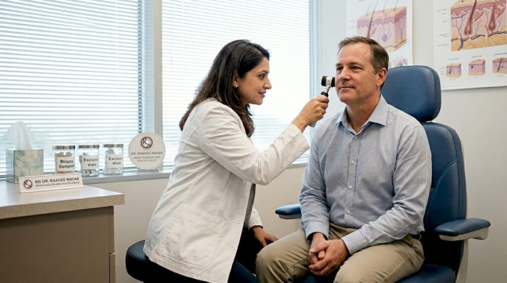
Once SCC is suspected, the process of confirming and characterising it begins. A specialist will first examine the lesion closely, often with a dermatoscope, a handheld device that illuminates and magnifies the skin to reveal features invisible to the naked eye. Suspicion alone, however, is never enough to guide treatment.
Diagnosis is confirmed by biopsy. A small sample of tissue is removed under local anaesthetic and sent to a pathologist, who examines it under a microscope. Biopsy and high-risk feature assessment together determine not just whether SCC is present, but how aggressive it is likely to behave.
The staging process then follows a logical sequence:
- Determine whether the SCC is in situ or invasive. In situ means the abnormal cells are confined to the epidermis and have not yet broken through to deeper layers. Invasive SCC has crossed that boundary.
- Assess high-risk features. These include tumour diameter greater than 2 cm, depth beyond 2 mm, poor differentiation under the microscope, perineural invasion (spread along nerve sheaths), and location on the ear or lip.
- Evaluate regional lymph nodes. For high-risk lesions, imaging or sentinel node biopsy may be recommended to check whether cancer has spread.
- Consider distant spread. Rare in early SCC, but relevant for advanced or immunocompromised patients.
This thorough work-up directly shapes the treatment plan. A small, well-defined, low-risk SCC on the trunk is managed very differently from a large, recurrent, or poorly differentiated tumour on the nose. Accessing expert skin cancer treatment planning at this stage prevents under-treatment and unnecessary tissue loss in equal measure.
“The biopsy result is not the end of the diagnostic journey; it is the beginning of a precise, risk-stratified treatment plan.”
Treatment options: Why Mohs surgery leads for facial SCC
Once staging is complete, treatment selection depends on tumour characteristics, anatomical location, and the patient’s overall health. Several options exist, and each has its place.
| Treatment | How it works | Best suited for | Cure rate |
|---|---|---|---|
| Standard excision | Removes tumour with a margin of normal tissue | Low-risk SCC on trunk or limbs | ~92% |
| Mohs surgery | Layer-by-layer removal with same-day margin analysis | High-risk or facial SCC | 94 to 99% |
| Curettage and electrodesiccation | Scraping and burning the tumour | Small, superficial, low-risk lesions | Variable |
| Radiotherapy | Targeted radiation to destroy cancer cells | Patients unfit for surgery; adjuvant use | Variable |
| Topical/immunotherapy | Creams or systemic agents | In situ or metastatic disease | Varies widely |
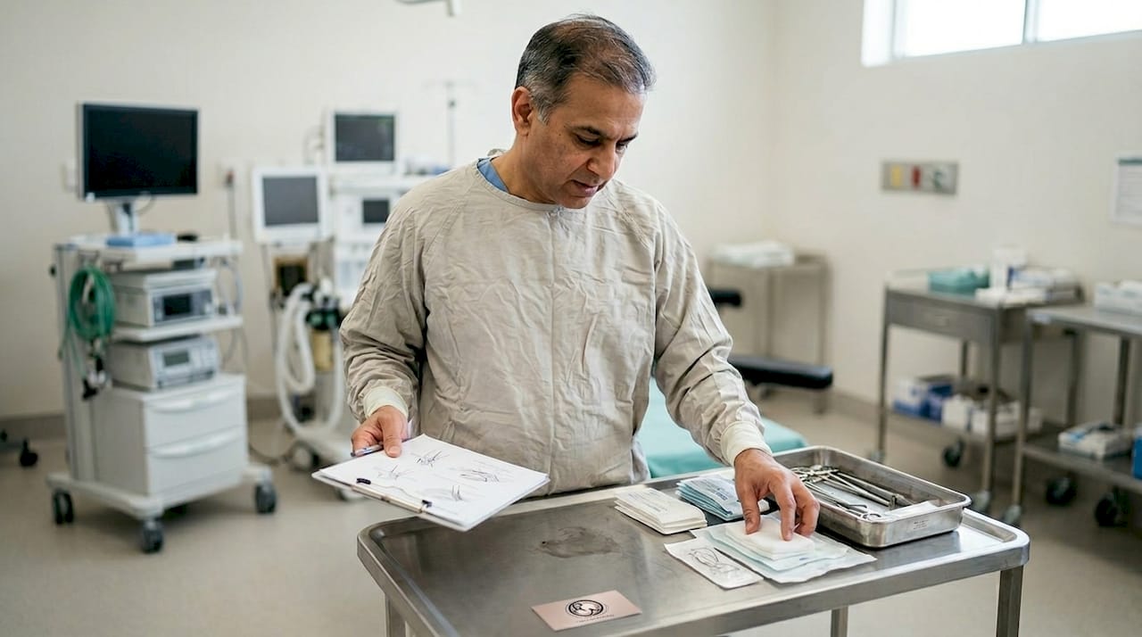
Mohs micrographic surgery is the gold standard for high-risk facial SCC, and the reason comes down to precision. During Mohs, the surgeon removes the visible tumour plus a thin rim of surrounding tissue, then immediately processes and examines 100% of the surgical margin under the microscope. If any cancer cells remain, only that precise area is removed next. This continues until the margin is completely clear.
The result is twofold. First, Mohs offers 94 to 99% cure rates, which are superior to standard excision. Second, because only cancerous tissue is removed, the defect left behind is as small as possible. On the face, where every millimetre matters, that tissue-sparing advantage is significant. Understanding what Mohs surgery involves before your appointment helps you feel prepared rather than anxious.
Mohs surgery is superior to excision for recurrent tumours and anatomically sensitive sites, including the nose, eyelids, lips, and ears. For patients with SCC on the nose, this distinction is particularly important given the nose’s complex three-dimensional structure.
Pro Tip: Ask your surgeon specifically whether your SCC meets the criteria for Mohs surgery. Not every SCC requires it, but for facial, recurrent, or high-risk tumours, it is rarely the wrong choice.
Facial reconstruction after Mohs: Balancing function and appearance
Removing the cancer is only part of the story. Once the tumour is clear, the surgical defect needs to be closed in a way that restores both function and appearance. This is where the skill of a surgeon trained in both Mohs and plastic surgery becomes genuinely transformative.
The goals of reconstruction are straightforward in principle but demanding in execution: restore normal facial movement and sensation, achieve a natural appearance, and tailor the approach to the individual patient’s anatomy, age, and lifestyle.
Surgeons choose from several techniques depending on the size, depth, and location of the defect:
- Primary closure: Direct stitching of wound edges, suitable for small defects with sufficient surrounding tissue
- Local flap: Adjacent skin is repositioned to cover the defect, closely matching texture and colour
- Skin graft: Skin taken from a donor site (often behind the ear) is placed over the defect when a flap is not feasible
- Secondary intention healing: The wound is left to heal naturally, appropriate for certain concave facial areas
Anatomical subunit preservation, such as keeping the nose and cheek as distinct units, is central to achieving a natural result. Flaps that respect these boundaries blend almost invisibly over time because they use skin of matching thickness and colour. Secondary intention healing is viable for select concave areas like the inner corner of the eye or the ear concha, where the natural contour actually aids healing.
The nose and cheek deserve special mention. The nose is the most common site for facial SCC, and its curved surfaces require careful planning. A detailed facial reconstruction guide explains the range of approaches available. For larger or more complex defects, facial reconstruction surgery may involve staged procedures to achieve the best possible result. Reviewing what reconstruction after Mohs typically involves, including timelines and what to expect, helps patients approach the process with realistic confidence. For nose-specific cases, dedicated information on reconstruction on the nose covers the nuances in detail.
Pro Tip: Before surgery, ask to see examples of reconstruction outcomes for your specific site. A surgeon experienced in facial artistry will welcome this conversation.
Nuances, limits and future directions for SCC management
Even with the best evidence and techniques, some aspects of SCC management remain genuinely complex. Large, recurrent, or aggressive tumours may require multiple Mohs stages in a single day, and certain anatomical locations demand particularly careful planning.
- Some facial SCCs require collaborative input from Mohs, reconstructive, and oncology teams
- Evidence for Mohs comes largely from cohort studies, not large randomised trials, though the data consistently shows superiority
- Emerging molecular therapies may one day allow personalised treatment targeting specific tumour mutations
- Ongoing monitoring after treatment is essential, as SCC carries a meaningful risk of recurrence or new primary tumours
Understanding reconstruction after Mohs and keeping up with your follow-up appointments are both part of managing SCC as a long-term condition rather than a single event. A broader skin cancer overview can help you understand where SCC sits within the wider landscape of skin cancer care.
“The best outcomes come from treating SCC as a team sport, not a solo procedure.”
Our perspective: What most people miss about SCC treatment
Most patients arrive focused on one question: will the cancer be gone? That is entirely understandable. But in our experience, the patients who feel most satisfied with their outcomes are those who also asked a second question: what will I look like afterwards, and how will I feel?
The distinction matters more than people realise. A surgeon who is expert at cancer clearance but less experienced in facial reconstruction may leave you with clean margins but an outcome that affects your confidence every time you look in the mirror. Conversely, prioritising cosmesis at the expense of thorough excision is equally problematic.
The real value of dual-trained expertise lies in holding both goals simultaneously, not trading one for the other. Aftercare and monitoring are equally underestimated. SCC patients have a higher lifetime risk of further skin cancers, and a structured follow-up plan is not optional. Reviewing realistic Mohs outcomes honestly, including what healing looks like week by week, prepares you far better than generic reassurance. Future-proofing your skin health is as important as the surgery itself.
Discover expert Mohs surgery and facial reconstruction options
If you or someone close to you is facing a diagnosis of squamous cell carcinoma, the quality of your surgical team matters enormously. Choosing a surgeon with dual training in both Mohs micrographic surgery and plastic surgery means your cancer clearance and your facial appearance are in the same expert hands.

Miss Rakhee Nayar offers precisely that combination, with specialist clinics in North West England and e-consultation options for patients across the UK and internationally. Whether you are seeking clarity on your diagnosis, exploring Mohs surgery explained, or ready to discuss expert facial reconstruction, the right guidance is available. Review all treatment options and take the next step with confidence.
Frequently asked questions
What does squamous cell carcinoma look like?
SCC often appears as scaly or nodular lesions on sun-exposed sites, presenting as a red scaly patch, a raised firm nodule, a non-healing sore, or a wart-like growth, most commonly on the face or ears.
How dangerous is squamous cell carcinoma?
With early treatment, SCC has a five-year survival rate of approximately 98%, but delaying treatment allows the tumour to grow deeper and increases the risk of spread to lymph nodes.
Why is Mohs surgery preferred for facial SCC?
Mohs excels for facial SCC because it combines the highest cure rates with precise tissue preservation, which is critical on the face where every millimetre of healthy skin affects both function and appearance.
What does recovery from facial SCC and Mohs look like?
Most patients experience excellent outcomes, with infection rates under 2% and reconstruction techniques that produce natural-looking results; full healing typically progresses over several weeks, with regular follow-up to monitor for recurrence.
Recommended
- Reconstruction After Mohs Surgery | Natural Results
- Miss Rakhee Nayar – Expert Mohs Surgery | UK
- What is Mohs Micrographic Surgery? | Miss Rakhee Nayar
- Skin Cancer Treatment & Surgery | Mohs Surgery UK
- How to Boost Skin Recovery Naturally for Radiant Results
Filed under Basal and Squamous Cell Carcinoma