TL;DR:
- Basal cell carcinoma can silently destroy facial tissues if untreated, despite rarely spreading distantly.
- Mohs surgery offers higher cure rates and tissue preservation, especially for facial high-risk BCCs.
- Proper post-surgical reconstruction ensures both functional and aesthetic facial outcomes.
Basal cell carcinoma is frequently dismissed as the “harmless” skin cancer. That label is dangerously misleading. While it is true that BCC rarely spreads to distant organs, it can silently destroy the nose, eyelid, or lip over months or years, leaving patients facing complex surgery they were never warned to prepare for. If your BCC is on your face, the stakes are higher than most people realise. Getting the right treatment, delivered by the right specialist, shapes not only your cure rate but also how you look and feel afterwards. This article walks you through what BCC actually is, how it is treated, and why the method and the surgeon both matter enormously.
Contents: Basal Cell Carcinoma, Mohs Surgery & Facial Reconstruction
- Understanding basal cell carcinoma
- Standard treatments for basal cell carcinoma
- When is Mohs surgery the preferred option?
- Aesthetic facial reconstruction after Mohs surgery
- A fresh perspective on BCC treatment: beyond the basics
- Explore expert Mohs surgery and facial aesthetic options
- Frequently asked questions
Key Takeaways
| Point | Details |
|---|---|
| BCC is common, not always mild | Basal cell carcinoma often develops on the face and can cause significant local damage if untreated. |
| Mohs surgery maximises cure and aesthetics | Mohs micrographic surgery is preferred for facial and high-risk BCCs for its high cure rate and tissue-sparing. |
| Expert reconstruction restores appearance | Advanced reconstructive techniques after Mohs help return natural facial contours and optimise cosmetic results. |
| Personalised care matters | Getting the right team for your BCC ensures both cancer removal and an aesthetic outcome tailored to you. |
Understanding basal cell carcinoma
Basal cell carcinoma begins in the basal cells, the deepest layer of your skin’s outer surface. These cells normally divide and replace older cells that shed from the surface. When UV radiation or other triggers cause genetic damage, some basal cells begin to divide uncontrollably, forming a slow-growing tumour that almost never spreads to other organs but is relentless in its local destruction.
BCC is, by a significant margin, the most frequent skin cancer worldwide. In the UK, it accounts for roughly 75% of all skin cancers diagnosed each year. Despite these numbers, many patients are still surprised to learn that it can affect critical cosmetic areas such as the face, nose, and eyelids, where treatment outcome impacts both cure and appearance.
Several key risk factors increase your likelihood of developing BCC:
- Cumulative UV exposure over a lifetime, particularly without sun protection
- Fair skin, light eyes, and red or blond hair, which offer less natural UV shielding
- History of sunburn, especially in childhood
- Immunosuppression, including long-term medication after organ transplant
- Radiation therapy to the skin in the past
- Family history of BCC or other skin cancers
Location changes everything with BCC. A small lesion on the back is a very different clinical problem from one sitting at the inner corner of the eye or along the nasal groove. The face contains structures that are cosmetically and functionally irreplaceable. Destroy the wrong millimetre of tissue and you may lose adequate eyelid closure, nasal airway function, or lip symmetry.
BCC often presents as a pearly or translucent pink bump, sometimes with visible blood vessels on its surface. It can also appear as a flat, scar-like lesion or a persistently crusting pink patch. Unlike melanoma, it rarely appears dark brown or black.
“If a spot on your face has been present for more than a few weeks, is not healing, and keeps returning after you scratch it, please have it assessed. Early diagnosis makes treatment far simpler and preserves far more tissue.”
When left untreated, BCC slowly invades cartilage, muscle, and even bone. The longer it remains, the more tissue must be removed to clear it, and the more complex the subsequent reconstruction becomes. Learning to recognise it early is essential; our guide to identifying BCC on the face outlines the key features to watch for.
Standard treatments for basal cell carcinoma
Not every BCC is treated in the same way. The right treatment depends on where the lesion sits, how large it is, what subtype it is, and whether it has ever been treated before. The main options currently available include:
- Curettage and cautery: scraping and burning the tumour, suitable for small, superficial BCCs on the trunk
- Standard surgical excision: cutting out the lesion with a margin of healthy tissue
- Radiotherapy: using targeted radiation, often reserved for patients who cannot tolerate surgery
- Topical treatments: creams such as imiquimod or 5-fluorouracil for superficial, low-risk lesions
- Mohs micrographic surgery: staged surgical removal with complete margin assessment
The table below illustrates how the two main surgical approaches compare:
| Feature | Standard excision | Mohs surgery |
|---|---|---|
| Margin assessment | Partial (breadloaf method) | 100% margin examination |
| Tissue conservation | Moderate | Maximum |
| 5-year cure rate | 90% primary, 94% recurrent | 97 to 99% |
| Best suited to | Small, low-risk, body BCCs | Face, high-risk, recurrent BCCs |
| Same-day reconstruction | Variable | Usually possible |
Standard excision for skin cancer is an excellent choice for straightforward, well-defined BCCs on areas where removing a wider margin is safe and uncomplicated. The problem arises when this approach is applied to the nose, eyelid, or ear, where a few extra millimetres of unnecessary removal can cause significant functional and cosmetic loss.
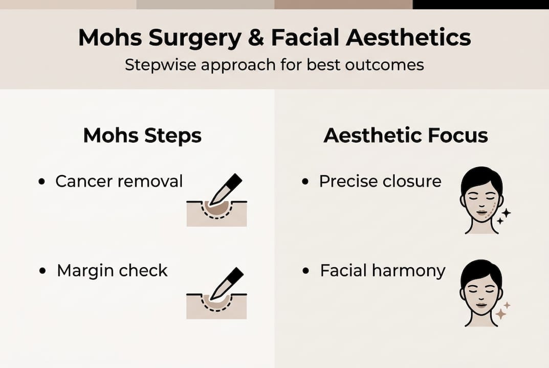
Mohs surgery, by contrast, examines every single edge and base of the tissue removed. The surgeon removes tissue in thin layers, maps each piece, and processes it in a controlled way to check all margins under the microscope while you wait. If any margin shows remaining cancer, only that specific area is addressed. This is how Mohs micrographic surgery achieves its superior cure rate while conserving the maximum amount of healthy tissue.
Pro Tip: Do not let anyone tell you that “quick removal” is always better for a facial BCC. Speed is irrelevant if the margins are not clear. An incomplete excision often means a second, more complicated procedure on an already-scarred area.
When is Mohs surgery the preferred option?
Mohs surgery is not necessary for every BCC. Its real strength lies in specific clinical situations where precision matters more than speed or simplicity. The following are the clearest indications for choosing Mohs:
- Location on the H-zone of the face: the central strip covering the eyelids, nose, lips, ears, and temples
- Tumour size greater than 1 cm in high-risk areas
- Ill-defined or poorly visible borders where standard excision cannot reliably judge margins
- Recurrent BCC, meaning a lesion that has come back after a previous treatment
- Aggressive histological subtypes such as morphoeic, infiltrating, or micronodular BCC
- Immunocompromised patients where higher precision reduces the risk of needing repeat surgery
The H-zone matters because these structures have no spare tissue. When treating Mohs surgery for BCC in the inner canthus of the eye, for example, even a millimetre of unnecessary loss can impair tear drainage and cause permanent discomfort.
The consequences of choosing a lesser technique for high-risk lesions are quantifiable. Aggressive BCC treated with curettage carries a 27% recurrence rate. Recurrent BCCs are harder to treat, require wider excision, and tend to present with more difficult reconstructive challenges.
At the same time, Mohs is sometimes applied to low-risk BCCs where it offers little added benefit over standard excision. Our high-risk skin cancer guide helps clarify which lesions genuinely warrant Mohs assessment.
Pro Tip: If your BCC sits near your eye, nose, or lip, always ask whether Mohs surgery has been considered. The answer to that question tells you a great deal about how thoroughly your treatment has been planned.
Understanding common misconceptions about this procedure is equally important. The Mohs surgery myths and facts guide addresses the most frequent concerns patients raise before their first consultation.
Aesthetic facial reconstruction after Mohs surgery
Clearing the cancer is only the first part of treatment. Once all margins are confirmed clear, the wound left behind must be repaired in a way that restores both function and appearance. This is where reconstructive expertise becomes just as critical as surgical precision.
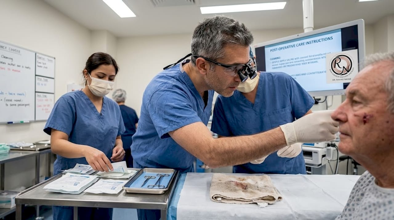
Facial reconstruction after Mohs surgery is guided by the subunit principle: the face is divided into aesthetic units (forehead, nose, cheeks, lips, eyelids), each with characteristic skin colour, texture, and thickness. Ideally, any repair uses tissue from within the same subunit or a closely adjacent one, because skin from a distant site often looks noticeably different once healed.
The approach varies by defect size and location:
| Defect size | Preferred closure | Typical location |
|---|---|---|
| Small (under 1 cm) | Direct linear closure | Cheek, forehead |
| Moderate (1 to 3 cm) | Local flap (e.g. bilobed flap) | Nose, temple |
| Large or complex | Skin graft or regional flap | Eyelid, lip, ear |
Local flaps are the workhorse of reconstruction after Mohs surgery. The bilobed flap, for instance, is specifically designed for nasal defects. It redistributes nearby skin using two rotations, keeping the repair within the same aesthetic subunit and avoiding the patchwork appearance that simpler methods can create.
Post-operative care is straightforward for most patients. Infection rates following Mohs with reconstruction are below 2%, and flap complications are uncommon when the procedure is performed by an experienced specialist. Swelling and bruising peak in the first week and settle considerably over four to six weeks, with final cosmetic results continuing to refine over several months.
“Patients consistently report that the anxiety before reconstruction is greater than the reality of recovery. Most find the process far less disruptive to daily life than they had anticipated.”
What produces the best outcomes is a team in which the Mohs surgeon and the reconstructive specialist are not two separate appointments but one integrated approach. This is the model used in facial reconstruction surgery that combines micrographic precision with genuine plastic surgery expertise.
A fresh perspective on BCC treatment: beyond the basics
One thing our clinical experience consistently reveals is that treating BCC is never simply a matter of removing a lump. Context shapes every decision. A 4 mm superficial BCC on the upper back is not the same clinical problem as a 9 mm morphoeic lesion on the alar rim of the nose, yet patients sometimes receive equivalent levels of urgency for both.
The risk of undertreating high-risk facial BCCs is well documented. But there is an equally real risk of overtreating low-risk lesions with techniques they do not need. Mohs is occasionally applied to straightforward lesions where standard excision would have been entirely adequate, adding cost and complexity without improving outcome.
For genuinely complex cases, including locally advanced BCC or lesions close to critical structures, the conversation must extend beyond surgery. Advanced imaging, hedgehog pathway inhibitors, and multidisciplinary input are all part of modern management. Our view at this practice is that the patient journey must integrate cure, cosmetic outcome, and emotional wellbeing together, not as sequential afterthoughts. Addressing Mohs misconceptions early in the consultation process helps patients arrive at decisions feeling informed rather than pressured.
Explore expert Mohs surgery and facial aesthetic options
If you have been diagnosed with a basal cell carcinoma on your face or another cosmetically sensitive area, you deserve care that treats the cancer and protects what matters to you aesthetically.

Miss Rakhee Nayar holds dual training in both Mohs micrographic surgery and plastic surgery, a rare combination that means diagnosis, removal, and reconstruction are handled with seamlessly integrated expertise. Whether you need expert facial reconstruction surgery following a previous diagnosis or are considering your treatment options for the first time, a personalised consultation will clarify exactly what approach is right for your specific lesion. Book your assessment through expert Mohs surgery and take the next step with confidence.
Frequently asked questions
Can basal cell carcinoma spread to other parts of the body?
BCC very rarely metastasises to distant organs, but untreated lesions cause significant local tissue destruction, making early treatment essential regardless of spread risk.
Why is Mohs surgery preferred for facial BCC?
Mohs surgery is preferred for facial BCC because it examines 100% of margins, removing only cancerous tissue while conserving healthy facial structures needed for optimal cosmetic reconstruction.
What is the recovery time after Mohs and facial reconstruction?
Most patients return to normal activities within a week, though cosmetic results improve gradually over several months as swelling resolves and tissue settles.
Are there risks or side effects after Mohs and reconstruction?
Infection occurs in under 2% of cases, and flap complications are uncommon, particularly when surgery is performed by a specialist with dedicated reconstructive experience.