
Skin flap reconstruction is defined as a surgical technique in which skin and underlying tissue are lifted from an area adjacent to a wound, moved to cover the defect, and kept attached to their original blood supply throughout. This approach is most commonly used after skin cancer excision, particularly following Mohs micrographic surgery, when the resulting wound is too large or too complex to close with simple stitches. Unlike a skin graft, a flap carries its own circulation, which gives it a significantly higher chance of surviving in its new position. Rakhee Nayar – Mohs Surgeon and Skin Specialist, a GMC-registered Consultant Plastic Surgeon with dual training in Mohs surgery and plastic surgery, uses flap techniques routinely to restore both function and appearance after cancer removal on the face and other cosmetically sensitive areas.
What is skin flap reconstruction and how does it differ from a graft?
Skin flap reconstruction is the standard reconstructive option when a wound cannot be closed directly and requires tissue with its own intact blood supply to heal reliably. The Northern Care Alliance NHS Foundation Trust describes the technique as skin origami, because the surgeon folds and rearranges adjacent normal skin to cover a defect while keeping the blood vessels connected.
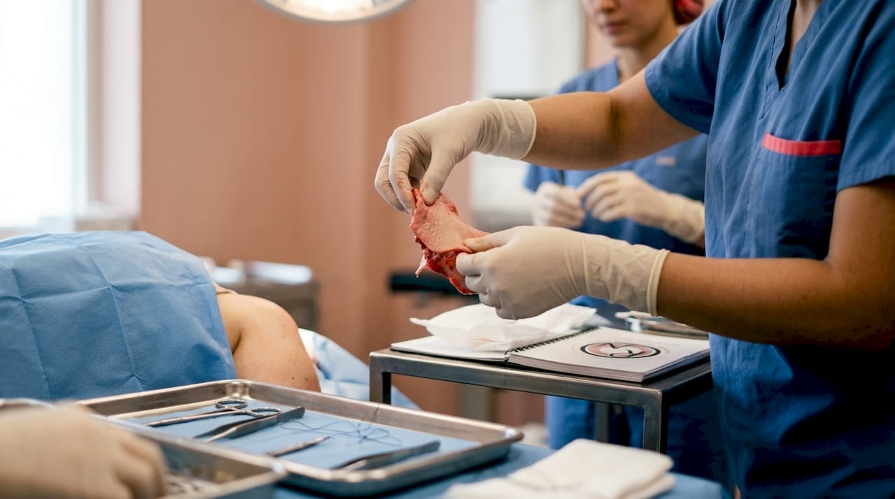
A skin graft, by contrast, is a sheet of skin cut entirely free from its donor site and placed over the wound. It has no blood supply of its own at the point of transfer. The graft must grow new connections from the wound bed beneath it, a process called inosculation, which takes longer and is less reliable on areas with poor circulation or irregular contours.
The critical advantage of a flap is that blood supply is preserved, making it more likely to heal quickly and survive in its new position. This matters most on the face, where the nose, eyelids, and lips have complex three-dimensional shapes that a flat graft cannot reproduce well. Flaps also tend to match the surrounding skin in colour and texture more closely, because the tissue comes from the immediate neighbourhood of the wound.
Patients who have undergone surgical excision for skin cancer will recognise that the size of the defect left behind depends on how deeply and widely the cancer has grown. When that defect is large, on a cosmetically sensitive site, or over a structure such as cartilage or bone, a flap is the preferred closure method.
How does skin flap surgery work?
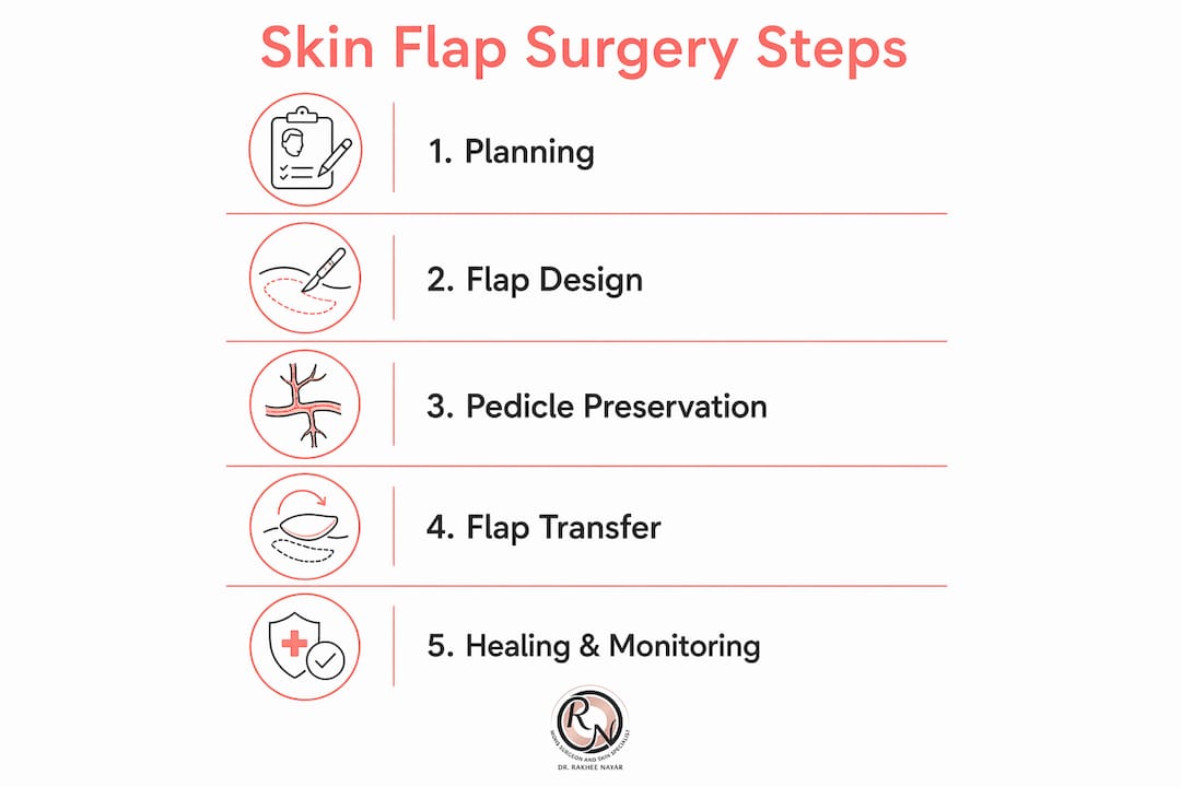
Skin flap surgery follows a logical sequence, and understanding each step helps patients know what to expect on the day of their procedure.
-
Marking the flap. The surgeon maps out a zone of adjacent skin large enough to cover the defect without placing the donor area under tension. The design accounts for the direction of natural skin creases, local blood vessel anatomy, and the need to hide scars in inconspicuous lines where possible.
-
Raising the flap. The skin and a layer of underlying fat are cut free on three sides, leaving one edge, called the pedicle, attached. The pedicle contains the blood vessels that keep the flap alive. The surgeon works carefully to protect these vessels throughout.
-
Moving the flap. Depending on the flap design, the tissue is advanced forward, rotated around a pivot point, or transposed across a short bridge of skin to reach the defect. Each movement type suits different wound shapes and locations.
-
Securing the flap. The flap is stitched into the defect. The donor site, the gap left where the flap was taken from, is closed directly with stitches or covered with a small skin graft if needed.
-
Applying dressings. A tie-over dressing is placed over the flap to hold it firmly against the wound bed and prevent movement that could disrupt the new blood supply connections forming beneath.
The vascular pedicle is the single most important element of flap success. If it is twisted, compressed, or damaged, the tissue loses its oxygen supply and the flap fails. This is why surgeons plan the geometry of the flap with great care before making any incision.
Pro Tip: Ask your surgeon to explain which direction your flap will move and where the donor site scar will sit. Knowing this in advance helps you understand what the wound will look like in the first two weeks, before swelling settles.
What are the types of skin flaps used in reconstruction?
Flap classification is based on two things: where the tissue comes from, and how it moves to reach the defect. Most flaps used after skin cancer excision on the face are local flaps, meaning the tissue comes from immediately beside the wound.
Local flaps
Local flaps are the most common choice for facial reconstruction after Mohs surgery. They use skin from the same cosmetic unit or an adjacent one, giving the best colour and texture match.
- Advancement flaps slide tissue directly forward into the defect along a straight line. They work well for elongated wounds on the forehead or scalp.
- Rotation flaps pivot a semicircular arc of skin around a fixed point to fill a triangular defect. They are frequently used on the cheek and scalp.
- Transposition flaps lift a rectangular or rhomboid segment of skin and swing it across a narrow bridge of intact skin to reach the wound. The rhomboid flap and the bilobed flap are two well-known examples used around the nose and temple.
Regional and distant flaps
Regional flaps borrow tissue from a nearby but not immediately adjacent area, such as the forehead flap used to reconstruct large nasal defects. Distant flaps, including free flaps transferred from the forearm or thigh with microsurgical vessel connections, are reserved for very large or complex defects and are rarely needed after standard skin cancer excision.
| Flap type | Typical site | Complexity | Healing considerations |
|---|---|---|---|
| Advancement | Forehead, scalp | Low to moderate | Minimal tension; linear scar |
| Rotation | Cheek, scalp | Moderate | Wider scar arc; good colour match |
| Transposition (rhomboid, bilobed) | Nose, temple, cheek | Moderate | Multiple incision lines; excellent contour |
| Forehead (regional) | Nose | High; two-stage | Staged division; longer recovery |
| Free flap (distant) | Large or complex defects | Very high | Microsurgery; hospital admission |
Choosing between these options depends on defect size, location, skin laxity, and the patient’s general health. Rakhee Nayar – Mohs Surgeon and Skin Specialist assesses each case individually, selecting the technique most likely to achieve both reliable healing and a natural appearance. Patients can read more about facial reconstructive surgery types to understand how these options apply to different areas of the face.
How is the skin flap healing process managed after surgery?
Healing after skin flap surgery follows a predictable pattern, though the timeline varies with flap size, location, and the patient’s general health. Most local flaps and the associated donor sites take 2–3 weeks to heal, with a specialist nursing review typically arranged at one week after the operation.
What to expect in the first two weeks
The first week is the most critical period for flap survival. The tissue may look bruised, swollen, or slightly darker than the surrounding skin. This is normal and reflects the adjustment of blood flow through the pedicle. Numbness around the flap is also common and usually resolves over several months as nerve endings regenerate.
- Keep dressings dry and undisturbed until the clinic review appointment.
- Avoid pressing, rubbing, or applying any product to the flap area.
- Sleep with your head elevated if the flap is on the face, to reduce swelling.
- Avoid strenuous activity, heavy lifting, and anything that raises blood pressure significantly for at least two weeks.
- Do not smoke. Nicotine constricts blood vessels and is one of the most significant risk factors for flap failure.
Stitches may be dissolvable or removable. Your surgical team will advise which type has been used and when any removable stitches will be taken out, usually at 7–14 days depending on the site.
Pro Tip: Photograph the flap at the same time each day in good natural light. If you notice a rapid change in colour, particularly a sudden darkening or pallor, contact your surgical team the same day. Early intervention can sometimes rescue a flap that is losing its blood supply.
Modern follow-up protocols such as Personalised Stratified Follow-Up (PSFU) tailor monitoring intensity to each patient’s individual risk profile. This means patients at lower risk of recurrence are not burdened with unnecessary clinic visits, while those at higher risk receive closer surveillance. The approach improves quality of life without compromising safety.
Wound care after facial surgery requires specific attention. Practical guidance on caring for facial surgery wounds covers dressing changes, cleaning, and signs that warrant urgent review.
What are the risks and potential complications of skin flap surgery?
Skin flap surgery is generally safe, but every surgical procedure carries risk. Understanding these risks helps patients make informed decisions and recognise early warning signs.
- Flap failure. Partial or complete loss of the flap occurs when blood supply to the tissue is insufficient. This is the most serious complication and is more likely in patients who smoke, have diabetes, or have significant cardiovascular disease.
- Infection. Any wound can become infected. Signs include increasing redness, warmth, swelling, and discharge from the wound. Antibiotics are usually effective when infection is caught early.
- Delayed healing. Some areas of the flap edge may be slow to heal, particularly at the corners of the repair where blood supply is most tenuous.
- Scarring. Because more incisions are required to free up the flap and close the donor site, flap surgery produces more scar lines than a simple direct closure. Scars typically fade and flatten over 12–18 months.
- Distortion of nearby structures. On the face, poorly planned or tensioned flaps can pull on the eyelid, nostril, or lip. Careful surgical planning minimises this risk.
- Haematoma. A collection of blood beneath the flap can compress the pedicle and threaten flap survival. Surgeons place drains or use pressure dressings to reduce this risk.
Patient factors that increase risk include smoking, uncontrolled diabetes, immunosuppression, and anticoagulant medication. Miss Nayar reviews all relevant medical history before surgery and may ask patients to stop certain medications or optimise their health in advance.
How does skin flap reconstruction compare to other reconstruction options?
Three main options exist for closing a wound after skin cancer excision: direct closure, healing by secondary intention, skin grafting, and skin flap reconstruction. Each has a distinct role.
| Method | Blood supply | Typical healing time | Best suited to |
|---|---|---|---|
| Direct closure | Intact (wound edges joined) | 1–2 weeks | Small defects with sufficient lax skin |
| Secondary intention | Wound granulates from base | 4–12 weeks | Concave sites; selected scalp wounds |
| Skin graft | None at transfer; grows new supply | 3–6 weeks | Flat, well-vascularised wound beds |
| Skin flap | Maintained throughout | 2–3 weeks | Complex, large, or contour-critical defects |
Skin grafts are simpler to perform than flaps but produce a less reliable cosmetic result on the face. The graft often appears slightly different in colour and texture from surrounding skin, and it may contract over time, distorting nearby structures. Grafts are well suited to large, flat wounds on the scalp or limbs where colour match is less critical.
Secondary intention healing, where the wound is left to close naturally from its edges and base, works well in concave areas such as the inner corner of the eye or the ear bowl, where the natural contraction of healing tissue actually improves the final shape. It requires no surgery beyond the initial excision but demands careful wound care over several weeks.
Skin flap reconstruction is the preferred choice when the defect is large, sits over a mobile structure such as the eyelid or lip, or requires three-dimensional contour to look natural. The reconstructive options after skin cancer depend on the size and depth of the defect, its location, and the patient’s overall health and preferences. Rakhee Nayar – Mohs Surgeon and Skin Specialist discusses all available options at consultation so that patients can make a fully informed choice.
Key takeaways
Skin flap reconstruction is the most reliable method for closing complex skin cancer wounds because it transfers tissue with an intact blood supply, giving it a higher chance of survival and a better cosmetic result than a graft.
| Point | Details |
|---|---|
| Blood supply is the key difference | Flaps retain their own circulation; grafts must grow new connections from the wound bed. |
| Local flaps suit most facial defects | Advancement, rotation, and transposition flaps offer the closest colour and texture match. |
| Healing takes 2–3 weeks on average | A specialist nursing review at one week monitors flap viability and dressing condition. |
| Smoking is the leading modifiable risk | Nicotine constricts blood vessels and significantly increases the risk of flap failure. |
| PSFU tailors follow-up to individual risk | Personalised Stratified Follow-Up reduces unnecessary visits while maintaining recurrence surveillance. |
What I have learned from planning skin flap reconstruction for my patients
Patients often arrive at consultation expecting reconstruction to be the most frightening part of their skin cancer treatment. In my experience, the opposite is true. Once the cancer is removed, reconstruction is where we restore what the disease took away, and that process is almost always more straightforward than patients anticipate.
The most important thing I have learned is that surgical planning matters more than surgical technique. A well-designed flap in the wrong orientation, or one that places the donor scar in a conspicuous position, can produce a technically successful but aesthetically disappointing result. I spend considerable time at the planning stage, mapping skin tension lines, assessing local tissue laxity, and considering how the repair will look not just at two weeks but at two years.
I also want to address a misconception I hear regularly. Patients sometimes assume that a larger flap will always give a better result. This is not the case. The smallest flap that reliably covers the defect is usually the best choice, because it minimises scarring, reduces the risk of distorting nearby structures, and shortens recovery. Bigger is not better in reconstructive surgery.
Preparation matters too. Patients who arrive for surgery well rested, non-smoking, and with their blood pressure well controlled heal faster and with fewer complications. If you are a smoker, stopping at least four weeks before surgery makes a measurable difference to flap survival. I always discuss this honestly with patients, because the evidence is clear and the benefit to them is real.
Finally, do not underestimate the value of a surgeon with dual training in both oncological excision and reconstruction. When the same specialist removes the cancer and plans the repair in a single procedure, the decisions about margin width, defect shape, and closure are made together rather than sequentially. That integration produces better outcomes for patients.
— Miss Rakhee Nayar
Skin cancer excision and reconstruction with Miss Rakhee Nayar
Miss Rakhee Nayar – Mohs Surgeon and Skin Specialist offers integrated skin cancer treatment and facial reconstruction at her private clinic in North West England. Her dual training means that Mohs micrographic surgery and reconstructive planning are considered together from the outset, not as separate steps.

Patients receive a personalised consultation covering diagnosis, excision options, and the full range of facial reconstruction surgery techniques, including local flaps, regional flaps, and skin grafts where appropriate. Private consultations and e-consultations are available for UK-based and international patients. To discuss your diagnosis and reconstruction options with Miss Nayar, contact the clinic directly to arrange an appointment.
This article is for information only and does not constitute medical advice. Consult a GMC-registered specialist for advice specific to your condition.
FAQ
What is skin flap reconstruction used for?
Skin flap reconstruction is used to close wounds that are too large or complex to join directly after skin cancer removal. It is most commonly performed after Mohs micrographic surgery on the face.
How does a skin flap differ from a skin graft?
A skin flap keeps its own blood supply throughout the procedure, making it more likely to survive and heal well. A skin graft is cut entirely free and must grow new blood connections from the wound bed beneath it.
How long does it take to recover from skin flap surgery?
Most local flaps and donor sites heal within 2–3 weeks, with a specialist nursing review at one week after surgery to check the flap and change dressings.
What are the signs that a skin flap is failing?
A rapid change in the colour or size of the flap area is the key warning sign. If the flap becomes suddenly pale, dark, or significantly swollen, contact your surgical team the same day.
Does smoking affect skin flap surgery outcomes?
Smoking constricts blood vessels and is one of the most significant risk factors for flap failure. Stopping smoking at least four weeks before surgery measurably improves the chances of a successful outcome.