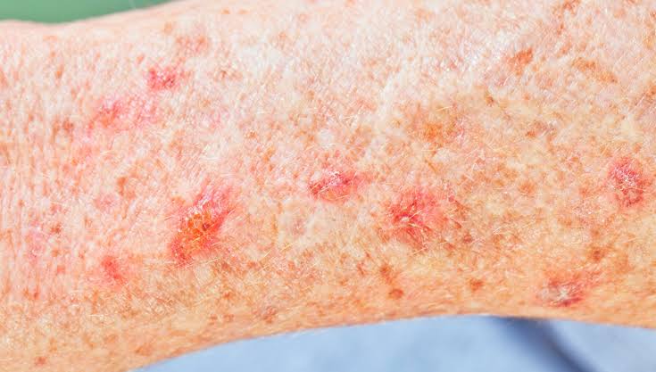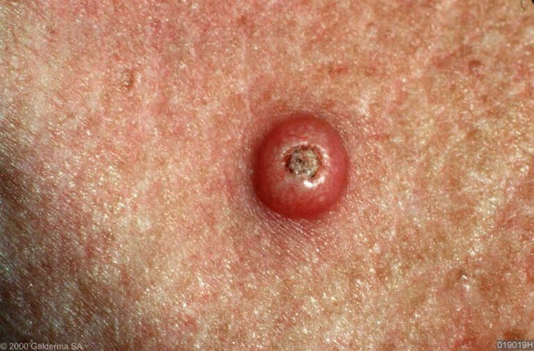
Melanoma in situ is defined as the earliest stage of melanoma, in which malignant melanocyte cells are confined entirely to the epidermis, the outermost layer of skin, without invading deeper tissue or spreading elsewhere in the body. Clinically classified as stage 0 melanoma, it carries an excellent prognosis precisely because it has not yet breached the basement membrane. The British Association of Dermatologists (BAD) and NICE both recognise it as a distinct and treatable condition. Understanding what is melanoma in situ, how it is detected, and what treatment involves gives patients the clearest possible foundation for informed decisions. This article draws on current UK clinical guidance and the expertise of Rakhee Nayar – Mohs Surgeon and Skin Specialist.
What causes melanoma in situ and how is it detected early?
Ultraviolet (UV) radiation is the primary cause of melanoma in situ. Cumulative sun exposure over decades, combined with episodes of sunburn, damages melanocyte DNA in the epidermis. This is why the condition appears most often in adults over 50, particularly on chronically sun-exposed sites such as the face, scalp, neck, and forearms.
Several risk factors increase the likelihood of developing the condition:
- Fair skin, light eyes, or red hair, which offer less natural UV protection
- A history of sunburn, especially in childhood or adolescence
- Multiple atypical moles (dysplastic naevi) on the body
- Previous skin cancer, including non-melanoma types
- Immunosuppression, whether from medication or illness
- Family history of melanoma in a first-degree relative
The early signs of melanoma follow the well-established ABCDE criteria: Asymmetry, Border irregularity, Colour variation (shades of brown, black, pink, or white within a single lesion), Diameter greater than 6mm, and Evolution or change over time. A lesion that itches, bleeds without trauma, or develops a new satellite patch warrants prompt assessment.
Epidemiological data shows that in situ melanoma subtypes are changing in England, with lentigo maligna rates stabilising while other subtypes continue to increase between 2013 and 2019. This trend underlines the importance of public awareness and routine skin checks.
Pro Tip: Photograph any changing mole monthly under consistent lighting. A simple side-by-side comparison over three months often reveals subtle colour or border changes that are easy to miss in real time.
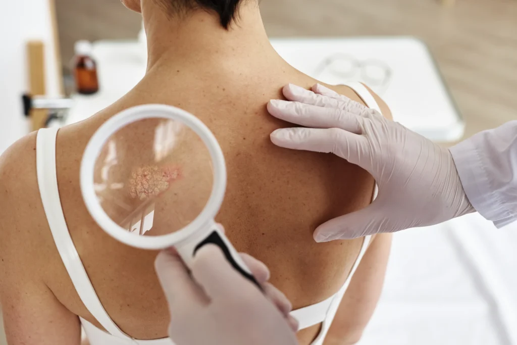
Dermoscopy, a non-invasive technique using a handheld magnifying device with polarised light, allows a trained clinician to examine pigment patterns beneath the skin surface. It significantly improves diagnostic accuracy compared with the naked eye alone. A biopsy remains the definitive next step when a lesion looks suspicious under dermoscopy.
How is melanoma in situ diagnosed?
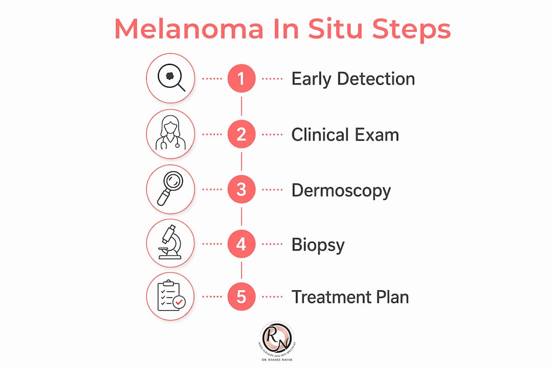
Diagnosis of melanoma in situ follows a structured pathway. A consultant first performs a clinical examination and dermoscopic assessment. If the lesion raises concern, a biopsy is taken for histological analysis.
Histology is the gold standard for confirming the diagnosis. A pathologist examines the tissue sample under a microscope and looks for specific features: atypical melanocytes arranged singly or in nests along the dermoepidermal junction, pagetoid spread of cells upward through the epidermis, and the absence of any dermal invasion. The presence of these features without breach of the basement membrane confirms the in situ diagnosis.
Key histological features that distinguish melanoma in situ from invasive melanoma and benign lesions include:
- Pagetoid spread: atypical cells scattered throughout the full thickness of the epidermis
- Confluent junctional nesting: clusters of melanocytes at the junction between epidermis and dermis
- No dermal invasion: the basement membrane remains intact
- Solar elastosis: surrounding skin often shows UV damage, particularly in lentigo maligna subtype
Reflectance confocal microscopy (RCM) is an emerging non-invasive imaging tool that can visualise cellular architecture in real time. It is used in specialist centres to map tumour margins before surgery, particularly for lentigo maligna on the face. RCM does not replace biopsy but reduces the number of mapping biopsies needed.
“Melanoma in situ is sometimes described as ‘pre-cancerous’ because it lacks metastatic potential, yet it contains genuinely malignant cells. That distinction matters clinically. It explains why treatment is still necessary, even though the prognosis is excellent. Patients who understand this framing tend to engage more constructively with their care.”
Adapted from European Society of Mohs Surgery clinical guidance on lentigo maligna and melanoma in situ
NICE and BAD guidelines both require histological confirmation before any definitive treatment begins. Clinical suspicion alone is not sufficient to proceed to surgery.
What are the treatment options for melanoma in situ?
Surgical excision is the standard treatment for melanoma in situ. The goal is complete removal of all malignant cells with a clear margin of surrounding normal skin. The specific approach depends on the lesion’s size, site, subtype, and the patient’s overall health.
Traditional wide local excision
Historically, UK guidelines recommended a 5–10mm clinical excision margin around the visible lesion. This approach is straightforward and widely available. For lesions on the trunk or limbs, it remains appropriate in most cases. The excised tissue is sent for histological analysis to confirm clear margins.
Tailored margin management
Recent UK centre policy changes reduce unnecessary wide local excisions by up to 70% for melanoma in situ by basing decisions on histological margin adequacy rather than fixed clinical distances. This shift is significant. It means patients with clear histological margins after an initial excision avoid a second, wider operation. The approach reduces surgical morbidity without compromising oncological safety.
The steps in a tailored excision approach typically follow this sequence:
- Initial diagnostic excision biopsy: the lesion is removed with a narrow margin to confirm diagnosis and assess histological clearance.
- Margin review by pathologist: the pathologist reports on the completeness of excision and any areas of concern.
- Multidisciplinary team (MDT) discussion: the case is reviewed by dermatology, pathology, and surgery together.
- Decision on further surgery: if margins are clear, no further excision is needed. If margins are involved, re-excision or specialist surgery is planned.
- Reconstruction if required: particularly for facial lesions, reconstruction is planned at the time of definitive excision.
UK guidelines prioritise shared decision-making and individualised management, with MDT involvement leading to bespoke treatment plans that account for patient comorbidities, tumour factors, and cosmetic considerations.
Mohs micrographic surgery for facial and recurrent lesions
Mohs micrographic surgery is preferred for lentigo maligna on the face or for recurrent lesions, due to better margin control and tissue conservation. In Mohs surgery, the tumour is removed in thin layers, with each layer examined under the microscope immediately. Surgery continues only until all margins are confirmed clear. This real-time margin assessment means the smallest possible amount of healthy tissue is removed.
For facial melanoma in situ, where cosmetic outcomes matter greatly, this approach is particularly valuable. Miss Nayar at Rakhee Nayar – Mohs Surgeon and Skin Specialist is dual-trained in both Mohs surgery and plastic surgery, which means reconstruction can be planned and performed at the same sitting. Patients benefit from latest Mohs surgery techniques that minimise scarring while achieving complete tumour clearance.
A comparison of the two main surgical approaches clarifies the decision:
| Feature | Wide local excision | Mohs micrographic surgery |
|---|---|---|
| Margin assessment | Post-operative (standard histology) | Intraoperative (real-time, 100% margin review) |
| Tissue conservation | Fixed margin removed regardless of tumour extent | Minimal healthy tissue removed |
| Best suited for | Trunk, limb, and straightforward lesions | Face, scalp, recurrent or large lesions |
| Reconstruction | Separate referral if needed | Planned at same sitting by dual-trained surgeon |
| Availability | Widely available in NHS and private settings | Specialist centres only |
Pro Tip: If your lesion is on the face, nose, eyelid, or ear, ask your dermatologist specifically whether Mohs surgery or slow Mohs is appropriate before consenting to standard wide local excision. The cosmetic difference can be substantial.
Non-surgical options such as imiquimod cream are occasionally used for patients who are unfit for surgery, particularly for large lentigo maligna lesions. These are not first-line treatments and carry a higher recurrence risk. Radiotherapy is another alternative in selected cases. Both are considered only when surgery is contraindicated.
What is the prognosis and what follow-up is recommended?
The prognosis after treatment for melanoma in situ is excellent. Because the malignant cells are confined to the epidermis and have not invaded the dermis or entered the lymphatic or vascular system, there is no risk of metastasis from the in situ lesion itself. This is the defining clinical advantage of catching melanoma at stage 0.
Patients generally require only one post-treatment check-up after melanoma in situ removal and typically need no routine follow-up or scans thereafter. Cancer Research UK guidance reflects the low recurrence risk after complete excision. This is reassuring for patients who may expect years of hospital appointments.
Recommended post-treatment steps include:
- One follow-up appointment at approximately 3 months to confirm wound healing and review histology
- Self-examination monthly: check the treated site and surrounding skin for any new changes
- Annual GP skin check if you have multiple risk factors, such as a history of atypical moles or previous skin cancer
- Sun protection year-round: SPF 30 or higher on all exposed skin, including on overcast days
- Vitamin D supplementation if sun avoidance is strict, as advised by your GP
The melanoma survival rate by stage confirms that stage 0 carries the highest survival figures of any melanoma classification. Patients treated with complete excision can reasonably expect no further melanoma-related illness from that lesion.
Recurrence is possible if the original excision margins were not clear, which is why histological confirmation of clearance is non-negotiable. Patients with lentigo maligna subtype on the face may have a slightly higher recurrence risk due to the subclinical spread of the lesion beyond its visible borders. This is precisely the scenario where Mohs surgery offers the greatest advantage.
After treatment, maintaining good skin health practices reduces the risk of developing a second primary melanoma. Patients with one melanoma in situ have a modestly elevated lifetime risk of developing another skin cancer, making ongoing skin awareness a permanent habit rather than a temporary precaution.
Key takeaways
Melanoma in situ is the most treatable form of melanoma because malignant cells remain confined to the epidermis, making complete surgical removal both achievable and curative in the vast majority of cases.
| Point | Details |
|---|---|
| Stage 0 melanoma | Malignant cells are confined to the epidermis with no invasion or metastatic risk. |
| Histology confirms diagnosis | Biopsy and pathological examination remain the only definitive diagnostic method. |
| Surgery is the primary treatment | Wide local excision or Mohs surgery, chosen based on site, size, and patient factors. |
| Mohs surgery for facial lesions | Preferred for lentigo maligna on the face due to real-time margin control and tissue conservation. |
| Minimal follow-up required | One post-treatment check is typically sufficient; no routine scans are needed after complete excision. |
Specialist care for melanoma in situ at Rakhee Nayar – Mohs Surgeon and Skin Specialist
Rakhee Nayar – Mohs Surgeon and Skin Specialist offers consultant-led assessment and treatment for patients diagnosed with or concerned about melanoma in situ, with private appointments available at Circle Cheshire in North West England and e-consultations for patients across the UK and internationally.
Miss Nayar’s dual training in Mohs micrographic surgery and plastic surgery means that diagnosis, excision, margin assessment, and reconstruction are managed by a single specialist. Patients with facial lesions benefit particularly from this integrated approach. For those who have received a biopsy result and are unsure of next steps, a skin cancer detection consultation provides clarity on diagnosis and a clear treatment plan. Patients seeking to understand their full range of options can also review the complete melanoma treatment guide on the website before their appointment.
Private consultation fees are available on request. This article does not constitute medical advice. Consult a GMC-registered specialist for assessment and treatment of any skin lesion.
FAQ
What is melanoma in situ in simple terms?
Melanoma in situ is skin cancer at its earliest stage, where abnormal melanocyte cells are present only in the outer layer of skin and have not spread deeper or to other parts of the body.
Is melanoma in situ serious?
It contains genuinely malignant cells and requires treatment, but its prognosis is excellent because it carries no risk of metastasis when completely removed.
What does melanoma in situ look like?
It typically appears as an irregular, flat patch with variable shades of brown, black, or pink, often with an uneven border. It may resemble a large freckle or a changing mole.
How is melanoma in situ treated in the UK?
Surgical excision is the standard treatment, with the approach tailored to the lesion’s site and size. Mohs micrographic surgery is preferred for facial or recurrent lesions, as confirmed by European and UK clinical guidelines.
Does melanoma in situ require long-term follow-up?
After complete excision, Cancer Research UK guidance indicates that one post-treatment check is typically sufficient, with no routine scans or long-term hospital follow-up needed in most cases.
Recommended
- Melanoma cancer treatment: your complete UK guide
- What is melanoma cancer: the expert guide for patients
- Melanoma mole: signs, risks, and treatment options
- Melanoma survival rate by stage: what patients need to know
Filed under Skin Cancer Explained