Skin cancer margins are defined as the border of normal-appearing skin removed alongside a tumour during surgical excision to confirm complete cancer removal. When a surgeon excises a skin cancer, the goal is not simply to remove the visible lesion. A rim of surrounding healthy tissue is taken at the same time, and this rim is the surgical margin. Pathologists then examine the edges of that removed tissue under a microscope to determine whether cancer cells reach the border. The result, reported as clear, close, or positive, directly shapes whether further treatment is needed. Guidelines from the British Association of Dermatologists (BAD) and NICE set the standard margin widths used across the UK, and Miss Rakhee Nayar, GMC-registered Consultant Plastic Surgeon and Mohs specialist, applies these in clinical practice at her North West England clinic.
What are skin cancer margins and why do they matter?
A surgical margin is the measured distance between the edge of a tumour and the cut edge of the excised tissue. Surgeons plan this distance before making any incision, using clinical assessment and established guidelines to decide how wide the margin should be. The margin serves one purpose: to reduce the chance that cancer cells remain in the body after surgery.
The importance of skin cancer margins lies in what happens when they are inadequate. If cancer cells reach the cut edge of the removed tissue, the pathologist reports the margin as “positive.” A positive margin means residual tumour may remain in the patient’s skin. That increases the risk of local recurrence and, in some cancers, spread to nearby lymph nodes or beyond.
Margin status is reported in three ways. A clear or negative margin means no cancer cells are seen at the tissue edge. A close margin means cancer cells are present but do not quite reach the edge, typically within 1 mm. A positive margin means cancer cells touch or breach the cut edge. Each status carries different clinical implications, which the treating team weighs carefully before deciding on next steps.
Understanding margin terminology helps patients read their pathology report with confidence. A clear margin is the goal of every excision, but it does not guarantee that no microscopic spread has occurred beyond the excised tissue. It confirms that the specimen itself appears free of tumour at its edges, which is the best available evidence of complete local removal.
How does margin width vary by skin cancer type?
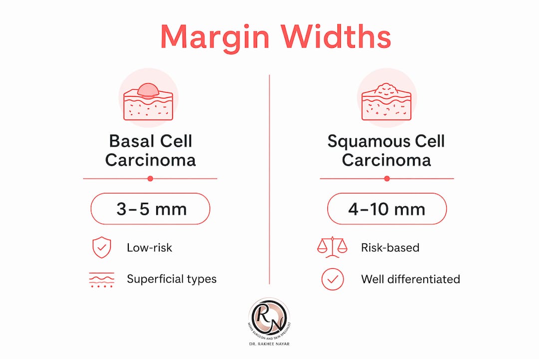
Margin widths vary by tumour type, with basal cell carcinoma (BCC) typically requiring 3–5 mm, squamous cell carcinoma (SCC) requiring 4–10 mm based on risk, and melanoma margins determined by Breslow depth. These are not arbitrary numbers. Each figure reflects decades of clinical evidence on how far each cancer type tends to spread beyond its visible border.
Basal cell carcinoma margins
BCC is the most common skin cancer in the UK. Low-risk BCCs, such as superficial or nodular subtypes on the trunk or limbs, are typically excised with a 3–4 mm margin. High-risk subtypes, including morphoeic, infiltrative, or recurrent BCCs, require wider margins of 5 mm or more because their growth pattern is less predictable and they spread along tissue planes that are invisible to the naked eye.
Squamous cell carcinoma margins
SCC margin planning depends on risk stratification. Low-risk SCCs, defined by small size, well-differentiated histology, and location on non-specialist sites, are typically excised with a 4–6 mm margin. High-risk SCCs, including those larger than 2 cm, poorly differentiated tumours, or those arising on the ear, lip, or in immunosuppressed patients, require margins of 6–10 mm. The BAD and NICE guidelines both support this risk-based approach.
Melanoma margins and Breslow depth
Melanoma margin planning is directly tied to Breslow depth, which measures the vertical thickness of the tumour in millimetres. The standard UK recommendations are:
- In situ melanoma: 5 mm margin
- Breslow depth up to 1 mm: 1 cm margin
- Breslow depth 1.01–2 mm: 1–2 cm margin
- Breslow depth greater than 2 mm: 2 cm margin
Breslow depth is the single most important prognostic factor in primary melanoma. A thicker tumour carries a higher risk of spread, which is why the margin widens proportionally.
Pro Tip: Ask your surgeon to confirm which subtype of skin cancer you have before surgery. The subtype, not just the diagnosis, determines the correct margin width. A nodular BCC and a morphoeic BCC are treated very differently.
| Cancer type | Standard margin | Key variable |
|---|---|---|
| Low-risk BCC | 3–4 mm | Subtype and site |
| High-risk BCC | 5 mm or more | Infiltrative growth pattern |
| Low-risk SCC | 4–6 mm | Differentiation and size |
| High-risk SCC | 6–10 mm | Size, site, immune status |
| Melanoma in situ | 5 mm | Confirmed in situ status |
| Invasive melanoma | 1–2 cm | Breslow depth |
How are skin cancer margins marked and assessed?
Accurate margin marking is a clinical skill that precedes the first incision. The process follows a defined sequence, and each step exists for a specific reason.
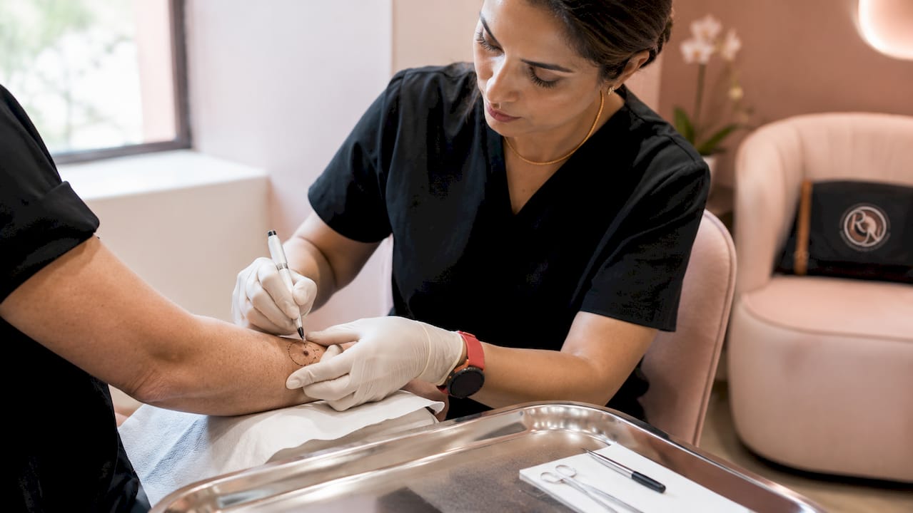
Clinical assessment before anaesthetic. Margin marking is done before anaesthetic injection to avoid tissue distortion that compromises visualisation. Local anaesthetic causes the skin to swell and blanch, which can obscure the tumour border. Marking first preserves the surgeon’s view of the true lesion edge.
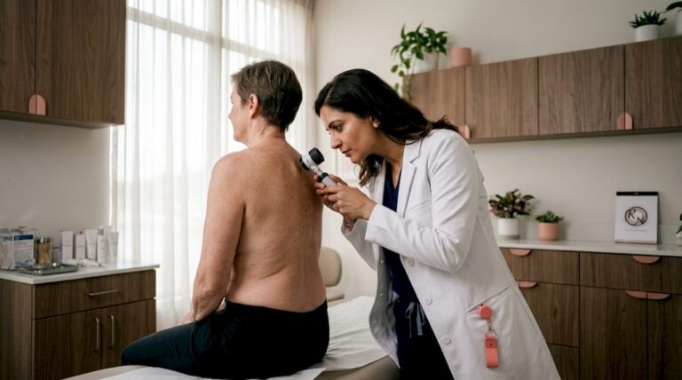
Use of magnification tools. Dermatoscopes and surgical loupes improve the accuracy of clinical margin assessment. A dermatoscope illuminates subsurface structures and helps identify tumour extension that is not visible to the naked eye. Loupes provide magnification during the excision itself, supporting precise cutting along the planned margin line.
Distinguishing diagnostic from therapeutic excision. Diagnostic excisions use smaller margins to confirm a diagnosis, whereas therapeutic excisions aim for wider margins to achieve complete removal. If a biopsy has already confirmed the cancer type, the surgeon plans a therapeutic excision with the full recommended margin from the outset.
Frozen-section analysis and Mohs micrographic surgery. In standard excision, the specimen is sent to a laboratory and results return within days. Mohs micrographic surgery takes a different approach. The surgeon removes the tumour with a thin margin, maps the tissue, and examines 100% of the cut edge under a microscope while the patient waits. If cancer remains, another layer is taken from the precise location of the positive margin. This continues until the margin is clear. Mohs surgery provides microscopic margin control that standard excision cannot replicate, which is why it is preferred for high-risk or cosmetically sensitive sites.
Pro Tip: If your skin cancer is on the face, eyelid, nose, ear, or lip, ask specifically whether Mohs micrographic surgery is appropriate for your case. These sites demand both maximum cancer clearance and tissue conservation, which is exactly what Mohs is designed to deliver.
What do margin pathology results mean for your treatment?
The pathology report is the document that tells you and your surgeon whether the excision achieved its goal. Clear margins mean no tumour cells are seen at the tissue edge; positive margins mean tumour cells reach or breach the cut edge. The distinction between these two results drives the next clinical decision.
When re-excision is recommended
Positive margins are the most common reason for a second operation. Margin positivity requiring re-excision occurs in 5–15% of cases, with lower rates seen in Mohs micrographic surgery. That figure reflects the reality that even experienced surgeons cannot always see every microscopic tumour extension with the naked eye. Re-excision removes the tissue around the original scar to clear any residual cancer cells.
The decision to re-excise is not automatic. Re-excision balances margin status with tumour location, patient health, and risk-benefit analysis. A positive deep margin in a frail patient with a low-risk BCC on the back may be managed with close observation rather than further surgery. A positive lateral margin in a young patient with a high-risk SCC on the temple almost always warrants re-excision or Mohs surgery.
Close margins and observation
A close margin, where cancer cells are present but do not reach the cut edge, sits in a clinical grey area. The treating team considers the tumour type, the distance from the margin, the site, and the patient’s overall health. Some close margins are observed with regular skin checks; others prompt further treatment. This decision is made within a multidisciplinary team (MDT) framework, which is standard practice in UK skin cancer care.
What a clear margin does and does not mean
A clear margin confirms tumour-free edges microscopically but does not guarantee the absence of microscopic spread beyond the excised tissue. This is a distinction worth understanding. A clear margin is the best evidence of complete local removal, but it does not eliminate the need for follow-up. Patients with a history of skin cancer require regular surveillance, regardless of margin status.
How do margins affect prognosis and reconstruction planning?
Margin adequacy is directly linked to outcomes. Standard margins achieve 92–95% clearance and reduce recurrence risk by 17–22%. Those figures represent the benefit of following guideline-recommended widths rather than taking a conservative approach to spare tissue.
Planning reconstruction around margin requirements
Reconstruction planning cannot begin until the surgeon knows the margin is clear. This is one of the most important practical points for patients to understand. If a surgeon plans a flap repair before confirming clear margins, and the margins return positive, the reconstruction may need to be undone. Mohs surgery solves this problem by confirming clear margins on the day of surgery, allowing immediate reconstruction in the same sitting.
The width and depth of the excision determine which reconstructive options are available. A small excision with primary closure is straightforward. A wider excision may require a skin graft or a local flap, which uses adjacent tissue to cover the defect. Excision depth may extend to fascia or periosteum when tumour invasion is suspected or when deeper clearance reduces re-excision risk. Deeper excisions create larger defects that require more complex reconstruction, including regional or free flaps in some cases.
Miss Rakhee Nayar’s dual training in Mohs surgery and plastic surgery means she can assess margin requirements and plan reconstruction simultaneously. This integrated approach reduces the number of procedures a patient needs and supports the best possible cosmetic outcome alongside cancer clearance.
Pro Tip: Before your excision, ask your surgeon to explain both the planned margin width and the likely reconstruction. Understanding both together gives you a realistic picture of the procedure, the scar, and the recovery.
Key takeaways
Surgical margin status is the single most important factor determining whether skin cancer excision has achieved complete local removal and whether further treatment is required.
| Point | Details |
|---|---|
| Margin definition | The border of normal skin removed with a tumour to confirm complete excision. |
| Width varies by cancer type | BCC requires 3–5 mm, SCC 4–10 mm, melanoma 1–2 cm based on Breslow depth. |
| Marking before anaesthetic | Margins are marked before injection to prevent tissue distortion obscuring the tumour edge. |
| Positive margins need review | Re-excision occurs in 5–15% of cases; the decision depends on tumour type, site, and patient factors. |
| Mohs improves clearance | Mohs micrographic surgery examines 100% of the cut edge, reducing positive margin rates significantly. |
What I have learned from years of managing skin cancer margins
Patients often arrive at a consultation focused on the diagnosis and understandably anxious about the word “cancer.” What they rarely expect is that the margin, a few millimetres of tissue around the tumour, will become one of the most consequential decisions of their treatment. In my experience, the margin conversation is where clinical judgement matters most.
Guidelines from the BAD and NICE give us the framework, and I follow them. But guidelines cannot account for every variable. A morphoeic BCC on the inner canthus of the eye, a high-risk SCC on the pinna, a melanoma close to a vital structure: these cases require a surgeon to weigh oncologic safety against functional and cosmetic outcomes in real time. That is not something a protocol can do for you.
The question I am asked most often is: “If my margins are clear, am I cured?” The honest answer is that clear margins are the best outcome we can achieve surgically, and they significantly reduce recurrence risk. But skin cancer surveillance remains important after any excision. Patients who have had one skin cancer are at higher risk of developing another, and regular skin checks are part of long-term care, not a sign that something went wrong.
What I find most valuable in my practice is the combination of Mohs surgery and immediate reconstruction. Patients leave the operating room knowing their margins are clear and their wound is already repaired. That certainty matters enormously. It removes the anxiety of waiting for pathology results before planning the next step, and it means the reconstruction is designed around a confirmed clear defect rather than an estimated one. For facial skin cancers in particular, that difference in approach produces measurably better outcomes, both oncologically and cosmetically.
— Miss Rakhee Nayar
Expert skin cancer care at Rakhee Nayar – Mohs Surgeon and Skin Specialist
Understanding your surgical margins is the first step. Getting them right is the next one. Rakhee Nayar – Mohs Surgeon and Skin Specialist offers consultant-led assessment, Mohs micrographic surgery, and integrated facial reconstruction at a private clinic in North West England, with e-consultations available for patients across the UK and internationally.

Miss Nayar’s dual training in plastic surgery and Mohs surgery means that margin planning and reconstruction are considered together from the outset. Whether you have received a new diagnosis or are concerned about a suspicious lesion, the skin cancer detection service provides expert assessment and a clear treatment plan. For patients weighing their surgical options, the guide on Mohs vs standard excision explains how margin outcomes differ between approaches. Private consultations are available; contact the clinic directly to arrange an appointment.
This article is for educational purposes only and does not constitute medical advice. Consult a GMC-registered specialist for guidance specific to your diagnosis and circumstances.
FAQ
What does a clear margin mean on a pathology report?
A clear margin means no cancer cells are detected at the cut edge of the removed tissue. It is the desired outcome of skin cancer excision, though regular follow-up remains necessary.
What happens if my skin cancer margins are positive?
Positive margins indicate cancer cells reach the edge of the excised tissue, suggesting residual tumour may remain. Re-excision or Mohs micrographic surgery is often recommended, though the decision depends on tumour type, location, and patient factors.
How wide should skin cancer surgical margins be?
Margin width depends on cancer type and risk. BCC typically requires 3–5 mm, SCC 4–10 mm, and melanoma 1–2 cm based on Breslow depth, following BAD and NICE guidelines.
Is Mohs surgery better for achieving clear margins?
Mohs micrographic surgery examines 100% of the cut margin during the procedure, which reduces positive margin rates compared with standard excision. It is particularly recommended for high-risk or cosmetically sensitive sites such as the face.
Can reconstruction be planned before margin results are known?
Reconstruction is ideally planned after margin clearance is confirmed to avoid undoing a repair if re-excision is needed. Mohs surgery allows same-day margin confirmation and immediate reconstruction, which is one of its principal clinical advantages.