TL;DR:
- Melanoma, a highly aggressive skin cancer responsible for most skin cancer deaths, can metastasize rapidly, especially if not detected early. Risk factors include fair skin, extensive moles, family history, and certain lifestyle factors, while early detection relies on the ABCDE rule and routine skin checks. Treatment varies from surgical excision for early stages to immunotherapy and targeted drugs for advanced disease, emphasizing the importance of specialist care and prompt diagnosis.
Melanoma accounts for just 1% of skin cancers yet causes the overwhelming majority of skin cancer deaths. That imbalance is what makes it so important to understand. Unlike basal cell or squamous cell carcinoma, melanoma can spread rapidly to lymph nodes and organs, transforming a skin condition into a systemic illness within months. Whether you have just received a diagnosis, had a suspicious mole removed, or carry risk factors that keep you vigilant, understanding what melanoma cancer is, how it behaves, and what treatment looks like gives you the foundation to make informed decisions about your care.
Key Takeaways
| Point | Details |
|---|---|
| Melanoma origins | Melanoma arises from pigment cells and can develop on normal skin or in moles. |
| Recognising symptoms | Spot unusual or changing moles using the ABCDE rule for early melanoma detection. |
| Staging importance | Accurate staging with Breslow thickness guides prognosis and treatment choices. |
| Treatment advances | Early melanoma is highly curable by surgery; advanced stages benefit from immunotherapy. |
| Prevent and monitor | Limit UV exposure and perform regular skin checks to reduce risk and catch changes early. |
What is melanoma and who is at risk
Melanoma is a malignant tumour originating in melanocytes, the specialised cells responsible for producing melanin, the pigment that gives skin its colour. When melanocytes suffer DNA damage, usually from ultraviolet (UV) radiation, they can begin dividing uncontrollably. What makes melanoma so dangerous compared to other skin cancer types is its early capacity to invade surrounding tissue and travel via the lymphatic system or bloodstream to distant organs.
Melanoma commonly arises on normal-appearing skin or from a pre-existing mole, and whilst sun-exposed areas like the back, shoulders, and legs are the most frequent sites, it can develop virtually anywhere. This includes the mucous membranes of the mouth and genitals, beneath fingernails and toenails, and inside the eye. Those last locations are particularly easy to overlook during routine self-examination.
Understanding your personal risk is not a simple checklist exercise. Several factors interact:
- Fair skin, light eyes, and red or blonde hair increase UV sensitivity and melanoma risk considerably
- A history of blistering sunburns, particularly in childhood, raises lifetime risk significantly
- More than 50 common moles or any atypical (dysplastic) moles increase susceptibility
- Family history of melanoma in a first-degree relative raises your risk two to three times
- Older men face the highest overall incidence, though melanoma is also one of the most common cancers in adults aged 15 to 29
- Immunosuppression, whether from medication or illness, reduces the body’s ability to detect and destroy abnormal cells
- Previous melanoma raises the risk of a second primary tumour
Reviewing the broader picture of skin cancer risk factors can help you contextualise where melanoma sits among the various diseases that affect the skin.
Recognising melanoma: signs and symptoms
The ABCDE rule is the most widely used framework for identifying suspicious lesions early, and for good reason. Over 80% of melanomas are first noticed by patients or their family members using this approach. Learning it takes minutes and can make a meaningful difference.
- A: Asymmetry. One half of the mole does not match the other when divided down the middle.
- B: Border. The edges are ragged, notched, or blurred rather than smooth and well-defined.
- C: Colour. Multiple shades within a single lesion, including browns, blacks, reds, whites, or blues, are a warning sign.
- D: Diameter. Lesions larger than 6mm (roughly the size of a pencil eraser) warrant attention, though smaller melanomas do exist.
- E: Evolving. Any mole that changes in size, shape, colour, or begins to bleed, itch, or crust deserves urgent assessment.
Where the ABCDE rule falls short is with nodular melanoma. This faster-growing subtype can be small, symmetrical, and uniformly coloured, making it easy to dismiss. The “ugly duckling” sign is a useful supplement: if one mole simply looks different from all the others on your body, treat it as suspicious regardless of ABCDE criteria.
Melanoma also develops in sites people rarely examine. Acral lentiginous melanoma, which disproportionately affects people with darker skin tones, appears on the palms, soles, and under nails. Monitoring these areas during self-examination is essential but often overlooked. For a broader understanding of skin cancer detection methods, regular full-body checks by a specialist are worth incorporating into your routine.
Pro Tip: Photograph any suspicious moles with your phone at monthly intervals. Sequential images make it far easier to detect subtle changes in size or colour that the naked eye misses in real time.

How melanoma is staged and why it matters
Staging is how doctors translate biological measurements into prognosis and treatment decisions. For melanoma, the two most critical pathological findings are Breslow thickness (how deep the tumour extends into the skin, measured in millimetres) and the presence of ulceration (whether the overlying skin surface has broken down). Both are assessed from the excised tissue sample after surgical removal.
The staging process, based on AJCC classification guidelines, works as follows:
- Stage 0 (melanoma in situ): Abnormal melanocytes are confined to the epidermis (the outermost skin layer) and have not invaded deeper tissue.
- Stage I: Tumour thickness of 2mm or less, with or without ulceration. Prognosis is excellent at this stage.
- Stage II: Tumour is thicker than 2mm or shows ulceration. Still localised but considered higher risk for spread.
- Stage III: Cancer has spread to regional lymph nodes or nearby skin. Treatment becomes more involved.
- Stage IV: Distant metastasis has occurred, meaning the cancer has spread to organs such as the lungs, liver, or brain.
| Stage | Breslow thickness | Ulceration | Approximate 5-year survival |
|---|---|---|---|
| IA | ≤1mm | Absent | Over 99% |
| IB | ≤2mm | Present or absent | 97% |
| IIA | 2.01 to 4mm | Absent | 94% |
| IIB | 2.01 to 4mm | Present | 87% |
| III | Any | Any | 40 to 78% |
| IV | Any (metastatic) | Any | Approximately 35% |
Ulceration at any thickness worsens outlook, which is why two tumours of identical depth can carry meaningfully different prognoses. Accurate skin cancer mapping at diagnosis is therefore not a bureaucratic formality; it directly shapes how aggressively your medical team will act.
Pro Tip: Ask your surgeon or oncologist for the exact Breslow thickness and ulceration status from your pathology report. These numbers are yours to know, and they determine every subsequent decision in your care pathway.
Current treatment options for melanoma
Treatment follows staging closely. Early-stage disease is predominantly surgical; advanced disease increasingly relies on systemic therapies that work on a molecular or immune level.
- Wide local excision (WLE): The primary treatment for stages 0 through II. Surgeons remove the tumour with a margin of healthy surrounding tissue. The margin width depends on Breslow thickness, ranging from 5mm for in-situ disease to 2cm for thicker tumours. Early-stage melanoma treated this way achieves cure rates between 85% and 99%.
- Sentinel lymph node biopsy (SLNB): Recommended for tumours thicker than 0.8mm. A dye is used to identify the first lymph node that drains the tumour site. Sampling this node reveals whether cancer has spread regionally, which is critical staging information.
- Immunotherapy: Drugs such as pembrolizumab and nivolumab (checkpoint inhibitors) work by releasing the immune system’s natural brakes, allowing it to attack melanoma cells. They have transformed advanced disease outcomes, with response rates of approximately 40 to 57%.
- Targeted therapy: Approximately 50% of melanomas carry a BRAF gene mutation. Drugs like vemurafenib and dabrafenib specifically target this mutation, producing rapid tumour shrinkage in eligible patients.
- Radiotherapy: Not a first-line melanoma treatment but used to manage local recurrence or brain metastases in specific scenarios.
| Treatment | Best suited to | Key benefit |
|---|---|---|
| Wide local excision | Stage 0 to II | High cure rate, definitive |
| Sentinel lymph node biopsy | Stage IB to II (thick) | Accurate staging, guides next steps |
| Immunotherapy | Stage III to IV | Durable responses in many patients |
| Targeted therapy (BRAF) | Stage III to IV with BRAF mutation | Rapid initial response |
For information on melanoma treatment options or guidance on managing high-risk melanoma, specialist consultation makes a significant difference in how your care plan is structured from the outset.
Pro Tip: Before starting systemic therapy for advanced melanoma, ask for BRAF and PD-L1 testing if it has not already been performed. Knowing your tumour’s molecular profile determines which drugs are most likely to work for you specifically.
Preventing melanoma and monitoring your skin
Knowing how to prevent melanoma overlaps significantly with understanding its causes. UV exposure remains the single most modifiable risk factor for the majority of melanoma subtypes. However, some melanoma types, particularly acral lentiginous melanoma, are not linked to sun exposure at all, which is why prevention cannot be reduced to sunscreen alone.
Practical steps that meaningfully reduce your risk:
- Apply broad-spectrum SPF 30 or higher sunscreen daily, including on overcast days and through car windows
- Avoid sunbeds entirely. UV output from sunbeds can be 10 to 15 times stronger than midday summer sun
- Wear protective clothing including wide-brimmed hats and UV-blocking garments, especially between 10am and 4pm
- Seek shade during peak UV hours rather than relying solely on sunscreen
- Perform a monthly full-body self-examination in good lighting, using a handheld mirror for hard-to-see areas
- Schedule annual skin checks with a dermatologist or skin cancer specialist if you have multiple risk factors
- Examine palms, soles, between toes, under nails, and inside the mouth regardless of sun exposure habits
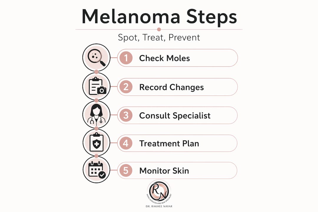
Monitoring melanoma prevention tips alongside routine early skin cancer detection strategies gives you the most complete protection currently possible.
Pro Tip: Keep a “skin diary” with dated photographs of any unusual spots. Bring this to every dermatology appointment. Visual history is often more valuable than memory alone when assessing whether a lesion has changed.
Rethinking melanoma care: expert insights and patient advocacy
Here is something statistics rarely communicate: being told you are in a “low-risk group” can be genuinely dangerous. Melanoma’s incidence is highest in older men, yet this demographic generalisation misleads younger people into complacency. A 24-year-old woman developing a melanoma on her lower leg will not find much comfort in population averages, and the clinical reality is that delays in diagnosis at any age tend to shift patients from an early treatable stage to a more complex one.
The other misconception worth confronting is that darker skin tones are protective against melanoma. They reduce UV-related risk considerably, but acral lentiginous melanoma shows no preference for skin tone. It affects the hands, feet, and nails equally across ethnicities. Because it develops in areas less routinely examined and is less associated with UV in public perception, it is frequently diagnosed later, and later diagnosis means worse outcomes.
Specialised care matters more than most patients realise at diagnosis. A general surgical excision performed without precise margin control can miss residual tumour tissue at cosmetically sensitive sites, leading to recurrence. Choosing a specialist skin cancer clinic with expertise in both oncological accuracy and reconstructive outcomes is not an indulgence. It is a clinical decision that directly affects long-term results.
The most empowered patients we see are not those who arrived with the most knowledge. They are the ones who came prepared with the right questions and a willingness to advocate for thorough investigation rather than a reassuring dismissal.
Expert support for melanoma treatment and skin cancer care
If you have received a melanoma diagnosis or carry significant risk factors, getting specialist input early shapes every decision that follows.

At mohssurgeon.co.uk, Miss Rakhee Nayar brings unique dual training in both plastic surgery and Mohs micrographic surgery to the management of skin cancer. Mohs surgery offers tissue-sparing, margin-controlled removal that is particularly valuable where preservation of function and appearance matter. Comprehensive skin cancer treatment services are available for a range of diagnoses and stages, with personalised care plans developed from your first consultation. For patients requiring restoration after excision, specialist facial reconstruction after skin cancer ensures that treatment outcomes support both your health and quality of life. In-person and e-consultations are available for UK-based and international patients.
Frequently asked questions
What exactly causes melanoma cancer?
Melanoma is mainly caused by DNA damage in pigment-producing cells from UV radiation exposure, but some types, like acral lentiginous melanoma, develop without sun exposure and affect all skin types equally.
How can I spot melanoma early on my skin?
Look for new or changing moles showing asymmetry, irregular borders, multiple colours, diameter larger than 6mm, or evolving characteristics; over 80% of melanomas are first spotted by patients or family using the ABCDE rule.
What are the main treatment options for melanoma?
Early melanoma is treated by surgical excision with 85 to 99% cure rates, while advanced melanoma may require immunotherapy or targeted drugs that achieve response rates of around 40 to 57%.
Is melanoma fatal or treatable?
Melanoma can be fatal when detected late, but localised melanoma carries over 99% five-year survival, making early detection the single most important factor in outcomes.
Who is at the highest risk for melanoma?
Older men face the highest overall melanoma risk, but the disease is also one of the most common cancers in young adults between 15 and 29, particularly women, making vigilance important across all age groups.
Recommended
- High-risk skin cancer: Expert treatment guide for UK patients
- Types of skin cancer and advanced facial treatments UK
- Understanding skin cancer mapping for precise treatment
- How to support skin cancer patients: practical help guide
Filed under Skin Cancer Explained