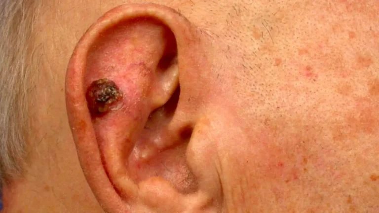
TL;DR:
- Early detection of suspicious skin patches is crucial for effective squamous cell carcinoma treatment.
- Typical SCC symptoms include non-healing sores, crusted patches, raised lumps, and bleeding ulcers.
- Diagnosis involves clinical exam, biopsy, and urgent referral if SCC is suspected.
Noticing an unusual patch of skin is unsettling, but the real difficulty lies in knowing whether it is something harmless or a sign of squamous cell carcinoma (SCC). Many UK residents dismiss rough, scaly patches as dry skin or an old scar, and that delay can matter. SCC is highly treatable when caught early, yet its symptoms overlap with dozens of ordinary skin conditions. This guide gives you a clear, evidence-based framework to assess what you are seeing, understand the full range of SCC symptoms, and know exactly when to act.
Key Takeaways
| Point | Details |
|---|---|
| Recognise core symptoms | Scaly patches, lumps, ulcers, or non-healing sores are most likely signs of SCC. |
| Early detection matters | Finding and addressing symptoms early increases treatability and reduces risk. |
| Risk varies by skin type | SCC symptoms and risks differ between fair skin and skin of colour—look beyond sun-exposed areas. |
| Seek urgent referral | Immediate GP review and urgent referral are recommended for suspicious symptoms. |
| Multidisciplinary care best | Advanced SCC requires coordinated care and surgery for optimal outcomes. |
How to assess skin changes for SCC: Initial criteria
Before you can interpret a specific symptom, you need a reliable way to evaluate whether any skin change warrants concern. Not every rough patch or new spot is dangerous, but certain features consistently separate suspicious lesions from benign changes.
Start by looking at four core characteristics: texture, colour, healing behaviour, and growth pattern. A normal spot of dry skin tends to improve with moisturiser within a week or two. An SCC lesion, by contrast, often persists, worsens, or changes in character over weeks and months. Colour shifts matter too. A lesion that transitions from pink to red, or develops a crusted, yellowish surface, deserves closer attention.
Size and depth add another layer. Lesions larger than 2 cm, or those that feel firm and anchored when you press gently, carry a higher risk profile. Location is equally important. Sun-exposed areas including the scalp, face, ears, lips, and backs of the hands are classic SCC sites, but lesions can also develop in areas that rarely see sunlight, particularly in people with certain risk factors.
SCC is the second most common skin cancer in the UK with over 45,000 new cases annually, and while most are treatable, a proportion can spread to lymph nodes or distant organs if left unaddressed. Knowing what to look for when spotting skin cancer symptoms on the face and elsewhere is the single most effective step you can take.
Key criteria to apply during a self-check:
- Persistence: Has the lesion been present for more than four weeks without improvement?
- Non-healing: Does it bleed, crust over, and then reopen repeatedly?
- Firmness: Does it feel indurated (hardened) compared to surrounding skin?
- Border irregularity: Are the edges poorly defined or raised unevenly?
- Tenderness: Is there pain or sensitivity without an obvious cause such as injury?
Pro Tip: Carry out monthly self-checks in good natural light using a hand mirror for hard-to-see areas. Do not limit checks to sun-exposed zones. Use the early detection guide to structure your self-examination routine.
The definitive squamous cell carcinoma symptoms list
With assessment criteria in mind, the next step is to lay out the definitive symptom list for SCC. These are the signs that clinicians look for, and they are the ones you should be able to name and describe when speaking to your GP.
Common symptoms of SCC include scaly or crusty patches, raised rough lumps, ulcers that bleed easily, non-healing sores, tenderness or pain, and firm indurated areas. These typically appear on sun-exposed sites such as the head, neck, ears, and hands.
Here is a structured breakdown of the core symptoms:
- Scaly or crusty patches: Often the earliest sign. The surface may look like dry skin but feels rough and adheres firmly to the underlying tissue.
- Raised, rough lumps: A dome-shaped or irregular bump that grows slowly over weeks. It may have a central depression or crater.
- Non-healing sores: A wound or ulcer that refuses to close over four to six weeks, even with standard wound care.
- Ulcers that bleed easily: Light contact causes bleeding. The base of the ulcer may appear red, moist, or necrotic.
- Tenderness or pain: Unlike many benign lesions, SCC can be genuinely painful, particularly when pressed.
- Firm, indurated areas: The lesion feels distinctly harder than surrounding skin, suggesting deeper tissue involvement.
- Wart-like growths: Some SCCs mimic viral warts, especially on the hands and fingers.
| Symptom | Typical appearance | Common location |
|---|---|---|
| Scaly patch | Rough, adherent scale | Scalp, face, forearms |
| Raised lump | Dome-shaped, irregular surface | Ears, lips, nose |
| Non-healing sore | Persistent ulcer, crusted edge | Hands, lower lip |
| Bleeding ulcer | Moist base, fragile surface | Face, neck |
| Indurated plaque | Hard, fixed to deeper tissue | Temples, cheeks |
SCC is highly treatable when diagnosed early, but it carries a real risk of metastasis in high-risk cases. Any non-healing lesion on sun-exposed skin lasting more than four weeks should be assessed by a clinician without delay.
Pro Tip: Do not assume a lesion is benign simply because it does not hurt. Early SCC is often painless. Pain tends to emerge as the lesion deepens or involves nerves. For a full overview of SCC diagnosis and surgery options, specialist assessment is the most reliable next step.
Symptoms in different skin types: What to look for
While most discussions of SCC centre on fair skin and sun exposure, it is vital to understand how the condition presents across different skin tones. Missing this distinction contributes to diagnostic delays that can have serious consequences.
In people with fair skin, SCC typically appears as a pink or red, scaly, raised lesion on sun-damaged areas. The contrast between the lesion and surrounding skin is usually obvious. In people with darker skin tones, the picture is more complex. In skin of colour, SCC is more common on non-sun-exposed areas such as the legs and anogenital region, tends to be potentially more aggressive, and diagnosis delays are more frequent. Understanding these SCC risks and genetics can help you advocate for timely assessment regardless of your skin tone.
| Feature | Fair skin | Skin of colour |
|---|---|---|
| Typical colour | Pink, red, or flesh-toned | Brown, dark, or hyperpigmented |
| Common location | Sun-exposed: face, ears, scalp | Non-sun-exposed: legs, genitalia, scars |
| Surface texture | Scaly, crusted | May appear smooth or verrucous |
| Aggressiveness | Variable | Often more aggressive |
| Diagnosis timing | Earlier on average | Frequently delayed |
Reasons why diagnosis may be delayed in people with darker skin tones:
- Lesions may not appear red or inflamed, making them less visually alarming.
- SCC arising in chronic scars or wounds (Marjolin’s ulcer) can be mistaken for normal scar tissue.
- Non-sun-exposed locations are less likely to be checked routinely.
- Awareness campaigns have historically focused on fair-skinned populations.
- Clinicians may have less experience recognising SCC presentations in darker skin.
Understanding these differences is not about creating alarm. It is about ensuring that everyone, regardless of skin tone, receives timely and accurate assessment.
Associated risk factors and early warning signs
Recognising what drives SCC risk, and which early signs matter most, helps you refine your personal assessment and decide how urgently to seek help.
UV exposure, immunosuppression, chronic wounds, and fair skin are the key risk factors, with organ transplant patients facing a particularly elevated risk due to long-term immunosuppressive medication. In fact, transplant recipients are up to 100 times more likely to develop SCC than the general population.
Precursor lesions are another critical consideration. Actinic keratosis (AK) and Bowen’s disease are pre-cancerous conditions that can progress to invasive SCC if left untreated. AK appears as rough, sandpaper-like patches on sun-damaged skin, while Bowen’s disease presents as a persistent, scaly, red plaque that is essentially SCC confined to the top layer of skin. Treating these precursors and addressing field cancerisation (widespread sun damage across a skin area) is an effective prevention strategy.
Statistical callout: SCC accounts for over 45,000 new diagnoses in the UK each year, making it the second most common skin cancer after basal cell carcinoma. The incidence is rising, partly due to an ageing population and cumulative UV exposure over decades.
Warning signs that should prompt immediate GP referral:
- A lesion that has not healed within four to six weeks
- Rapid growth of a lump or plaque over a matter of weeks
- A lesion that bleeds spontaneously or with minimal contact
- New or worsening pain around a skin lesion
- Swollen lymph nodes near a suspicious skin lesion
- A lesion arising within a longstanding scar or chronic wound
Knowing these triggers means you can act quickly rather than waiting for a routine appointment. Understanding follow-up after skin cancer treatment is equally important for those who have previously had a diagnosis.
What happens after noticing symptoms: Diagnosis and next steps
When symptoms or risk factors are present, knowing the diagnostic and treatment journey makes decision-making easier and reduces anxiety about the unknown.
The standard diagnostic pathway in the UK follows these steps:
- Clinical examination: Your GP or dermatologist examines the lesion visually, noting size, shape, colour, borders, and texture.
- Dermoscopy: A handheld device with magnification and polarised light allows the clinician to assess subsurface structures not visible to the naked eye.
- Biopsy: A small sample of tissue is removed under local anaesthetic and sent to a pathology laboratory. This is the only way to confirm SCC with certainty.
- Staging assessment: If SCC is confirmed, imaging such as ultrasound or CT scanning may be used to assess whether the cancer has spread to lymph nodes or beyond.
Diagnosis typically involves clinical examination, dermoscopy, and biopsy, with urgent referral on the two-week wait pathway if SCC is suspected. This NHS pathway means you should be seen by a specialist within two weeks of your GP making a referral.
Urgent referral indicators include:
- A lesion with features strongly suggestive of SCC on clinical examination
- A rapidly growing nodule or ulcer in a high-risk patient
- Any lesion in an immunosuppressed individual that does not resolve promptly
UK guidelines from NICE and the British Association of Dermatologists emphasise urgent referral and multidisciplinary care for advanced cases. Surgery remains the gold standard treatment for most SCCs, offering the highest cure rates and the most reliable margin assessment.
For lesions on the face or other cosmetically sensitive areas, the choice of surgical technique matters enormously. Mohs micrographic surgery offers real-time margin control, meaning the surgeon checks every edge of the removed tissue before closing the wound. This approach is particularly well-suited to SCCs on the face, ears, and scalp. A detailed surgical excision treatment guide can help you understand the options available and what to expect from each.
A specialist’s perspective: The real challenge in early SCC detection
In clinical practice, the cases that concern us most are not the obvious ones. A large, ulcerated lesion on the face of a fair-skinned patient with decades of sun exposure is rarely missed. The cases that slip through are the subtle ones: a slightly rough patch on the lower lip that a patient has attributed to chapping for six months, or a firm nodule on the ear that was assumed to be a cyst.
The most underappreciated aspect of SCC is the role of precursor lesions. Patients often receive treatment for actinic keratosis without fully understanding that these patches represent a genuine pre-cancerous process. When field cancerisation is present, meaning large areas of skin show cumulative UV damage, the risk of SCC developing somewhere within that field is substantial. Treating one lesion while ignoring the surrounding skin is like removing a single weed from a garden that is entirely overgrown.
Multidisciplinary assessment is not just for advanced cases. Even early-stage SCC on the face benefits from input that combines oncological precision with reconstructive expertise. The goal is not simply to remove the cancer. It is to remove it completely and restore form and function with the best possible outcome. Patients who seek specialist care early, before a lesion has grown or spread, consistently have better results, fewer complications, and greater peace of mind.
One misconception we encounter regularly is the belief that SCC only affects older, fair-skinned individuals with a history of outdoor work. In reality, SCC can arise in younger patients, in people with darker skin tones, and in areas that have never seen direct sunlight. Challenging these assumptions is part of why education matters. For a broader view of what is fact and what is fiction, the article on skin cancer myths addresses many of the beliefs that delay people from seeking help.
Pro Tip: At your next dermatology or GP appointment, ask specifically about field cancerisation and whether your sun protection routine is adequate for your individual risk level. This single conversation could prevent a future SCC.
Connect with expert care for SCC symptoms and treatment
If you have identified symptoms that concern you, the next step is to speak with a specialist who combines diagnostic precision with surgical expertise.

At mohssurgeon.co.uk, Miss Rakhee Nayar offers specialist assessment and evidence-based treatment for SCC, including Mohs surgery for lesions on the face and other sensitive areas. Her unique dual training in plastic surgery and Mohs micrographic surgery means you receive both the highest cure rates and the best possible aesthetic outcome. Whether you are seeking a second opinion, a private consultation, or comprehensive skin cancer treatment from diagnosis through to reconstruction, the clinic provides a clear, supported pathway. Private consultations and e-consultations are available for UK and international patients. Book your skin cancer diagnosis appointment today and take the most important step towards certainty.
Frequently asked questions
What are the most common symptoms of squamous cell carcinoma?
The most common symptoms are scaly or crusty patches, raised rough lumps, non-healing sores, ulcers that bleed easily, and tenderness, typically on sun-exposed skin such as the face, neck, ears, and hands.
How quickly should I see a doctor if I notice these symptoms?
See your GP without delay if you have a suspicious, non-healing, or painful skin change; urgent referral on the two-week wait pathway is recommended for any lesion where SCC is clinically suspected.
Are SCC symptoms different in skin of colour?
Yes. In skin of colour, SCC often presents on non-sun-exposed areas such as the legs or genitalia, may be more aggressive, and is frequently diagnosed at a later stage due to reduced awareness and atypical presentation.
What are the risk factors for developing SCC?
UV exposure, immunosuppression, chronic wounds, and fair skin are the primary risk factors, with organ transplant patients facing a significantly elevated lifetime risk due to long-term immunosuppressive therapy.
What treatments are available for SCC in the UK?
Surgery is the gold standard for most SCCs, with Mohs micrographic surgery offering the highest precision for facial and cosmetically sensitive lesions; treatment planning often involves multidisciplinary input for complex or advanced cases.
Recommended
- What Is Squamous Cell Carcinoma (SCC)? – Expert Mohs Surgery | UK
- About Basal Cell Carcinoma (BCC) – Expert Mohs Surgery | UK
- How to identify basal cell carcinoma on the face: UK guide
- Pictures of Basal Cell Carcinoma (BCC) – Warning Signs – Expert Mohs Surgery | UK
Filed under Spotting Skin Cancer