TL;DR:
- Mohs surgery offers high cure rates and precise tissue removal for high-risk basal cell carcinomas.
- The procedure involves staged excision, microscopic margin analysis, and immediate reconstruction options.
- Preparing thoroughly and understanding the process helps patients achieve better outcomes and confidence.
A diagnosis of basal cell carcinoma (BCC) rarely comes without a flood of questions. Where does the cancer end? Will surgery leave a mark? What actually happens in the operating room? Mohs micrographic surgery is widely regarded as the most precise method for removing BCC, particularly on the face, because it removes cancer layer by layer while examining every margin under a microscope. This guide walks you through every stage: from understanding why Mohs is chosen, through preparation, the surgical procedure itself, reconstruction, and recovery, giving you the clarity you need to face treatment with confidence.
Key Takeaways
| Point | Details |
|---|---|
| Precise cancer removal | Mohs surgery removes basal cell carcinoma with the highest precision, sparing healthy tissue. |
| Stepwise, on-site analysis | Stage-by-stage tissue checking ensures all cancer is cleared before wound closure. |
| Personalised reconstruction | The surgical site is repaired with the approach best matched to your location and needs. |
| Active patient role | Preparation, informed questions, and following aftercare instructions support your best healing. |
When is Mohs surgery used for basal cell removal?
Mohs micrographic surgery is not offered for every BCC. It is specifically selected when precision matters most, whether because of where the tumour sits on your body, how aggressive it is, or whether it has returned after previous treatment. As BAPRAS patient information confirms, Mohs is indicated for BCC on the head and neck, recurrent tumours, aggressive subtypes, ill-defined borders, and high-risk locations such as the nose, eyes, and lips where tissue and function must be preserved.
The logic is straightforward. Standard excision removes a margin of tissue around the tumour and sends it to a lab, where only a small percentage of the margin is actually examined. Mohs, by contrast, examines 100% of the surgical margin in real time. This is why it is used for lesions where incomplete removal would be dangerous or disfiguring. You can read more about Mohs micrographic surgery and why it is the preferred option for so many patients in the UK.
High-risk BCC features and locations that often warrant Mohs include:
- Tumours located on the nose, eyelids, lips, ears, or temples
- Recurrent BCC that has returned after prior treatment
- Aggressive histological subtypes such as morphoeic, infiltrative, or micronodular BCC
- Lesions with poorly defined clinical borders
- Large tumours or those close to critical structures
- BCCs in immunocompromised patients
- Any lesion where maximum tissue conservation is a clinical priority
The relationship between surgical precision and aesthetic outcome is particularly important on the face. Removing too little risks incomplete clearance; removing too much risks unnecessary functional or cosmetic impairment. This is the tension that Mohs resolves. You can explore how Mohs and facial aesthetics work together in specialist care.
Statistic: Studies consistently show Mohs achieves cure rates of up to 99% for primary BCC and approximately 94% for recurrent BCC, making it the gold standard for high-risk lesions.
What to expect on surgery day: preparation and prerequisites
Understanding why Mohs is used helps you appreciate the planning involved. The procedure itself is performed as a day case under local anaesthetic, meaning you are awake throughout and go home the same day. What surprises many patients is how long the day can feel. As NHS guidance confirms, each stage involves a period of waiting while the lab analyses your tissue, which can take up to two hours per stage, meaning you may be at the clinic for most of the working day.
The following checklist covers the essentials so you are fully prepared:
| Category | What you need to know |
|---|---|
| Items to bring | Snacks and drinks, a book or device for waiting, your medication list, reading glasses if needed |
| Clothing | Loose, comfortable clothing; avoid wearing make-up or products near the surgical site |
| Medications | Discuss blood thinners with your team in advance; most other medications continue as normal |
| Transportation | Arrange a lift home; do not drive after the procedure, especially if sedation is anticipated |
| Support person | Strongly advised to have someone with you; reconstruction may affect how you feel post-procedure |
| Fasting | Generally no fasting required for local anaesthetic, but follow your specific team’s instructions |
Your care team will also explain the following before the procedure begins:
- What local anaesthetic to expect and how it will be administered
- How the tissue specimen will be removed and mapped
- Approximately how many stages are anticipated
- What reconstruction is being considered and any alternatives
- When to expect your results and follow-up plan
Pro Tip: Bring a good book, a podcast, or something genuinely absorbing for the waiting periods. Most patients spend the majority of their day simply waiting between stages, and being mentally occupied makes the experience far more manageable.
A full walkthrough of preparation for Mohs surgery can help you feel entirely ready before the day arrives. It is also worth reading about the Mohs surgery for BCC step-by-step process so that nothing on the day comes as a surprise.
Step-by-step: Mohs micrographic surgery for basal cell removal
Being fully prepared for surgery day sets you up for a smooth procedure. Here is a transparent account of every step your specialist will take to ensure complete cancer removal.
The Mohs removal process involves three clearly defined steps, repeated as many times as necessary until a clear margin is confirmed:
-
Local anaesthetic and initial excision. The surgeon injects local anaesthetic around the tumour. Once the area is numb, the visible tumour plus a thin layer of surrounding tissue is removed. The specimen is then carefully mapped and colour-coded, so that any remaining cancer can be precisely located if it turns up under the microscope.
-
Laboratory analysis. The excised tissue is taken to an on-site lab, where it is frozen, sectioned, stained, and examined microscopically. This comprehensive margin examination looks at all surgical margins, not just a sample, which is what makes Mohs so accurate.
-
Additional targeted removal if needed. If cancer cells are still present at any margin, the surgeon removes another thin layer of tissue only from the precise area shown by the map to be affected. This cycle continues until margins are completely clear.
Typical timing: each surgical excision stage takes approximately 30 minutes, followed by up to two hours of laboratory analysis. Many patients are clear in one stage, but complex tumours may require two, three, or occasionally more stages.
Roughly half of all Mohs procedures achieve clear margins in a single stage. However, aggressive or larger BCCs may require additional passes. Each extra stage targets only the specific area of remaining cancer, which is how Mohs spares the maximum amount of healthy tissue.
| Feature | Mohs surgery | Standard excision |
|---|---|---|
| Margin examination | 100% of margin checked in real time | Approximately 1-5% sampled |
| Tissue conservation | Maximum healthy tissue spared | Wider safety margin routinely taken |
| Cure rate (primary BCC) | Up to 99% | Approximately 95% |
| Same-day result | Yes, confirmed before wound closure | No, results reported days later |
| Typical stages | 1-3 stages | Single excision |
| Best suited for | High-risk, facial, recurrent BCC | Lower-risk, non-facial tumours |
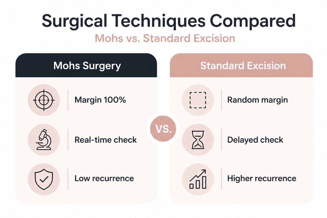
This comparison makes it clear why Mohs is not simply a more complex version of standard excision. It is a fundamentally different approach to achieving certainty.
Rebuilding and healing: reconstruction after basal cell removal
After confirming all cancer cells are cleared, the focus shifts entirely to healing. The good news is that in most cases, reconstruction options are discussed in advance and performed immediately after clear margins are achieved, on the same day as removal. There are five main approaches, and your surgeon will recommend the most appropriate based on the size and location of the wound, the quality of surrounding tissue, and your own preferences.
The five main reconstruction options are:
- Secondary intention healing. The wound is left to heal naturally without closure. This suits smaller wounds in certain locations and often produces very acceptable cosmetic results with minimal intervention.
- Primary closure. The wound edges are brought together and sutured directly. Simple, fast, and effective for smaller excision sites where there is sufficient tissue mobility.
- Local skin flap. Nearby skin is lifted and repositioned to cover the defect. This technique uses tissue that closely matches the colour and texture of the original area, making it particularly well suited to facial reconstruction.
- Skin graft. Skin is taken from a donor site elsewhere on the body and used to cover the wound. Grafts are often used for larger defects or areas where flap tissue is unavailable.
- Complex or staged repair. Some larger or more anatomically challenging wounds require multi-stage reconstruction, sometimes involving referral to a specialist reconstructive surgeon.
| Closure method | Pros | Cons | Recovery time |
|---|---|---|---|
| Secondary intention | No sutures, simple | Slower healing, open wound care | Weeks to months |
| Primary closure | Quick, straightforward | Requires tissue mobility | 1-2 weeks |
| Local flap | Excellent colour match, less visible scar | More complex procedure | 2-4 weeks |
| Skin graft | Covers large defects | Donor site needed, texture variation | 4-6 weeks |
| Complex repair | Best for challenging anatomy | Multiple stages, longer recovery | Variable |
Several factors influence the choice of approach:
- The anatomical location and how visible it is
- The size of the wound left after removal
- The condition and availability of surrounding tissue
- Your own health, healing capacity, and aesthetic priorities
Pro Tip: Ask your surgeon to share photographs of previous reconstructions in similar locations. Seeing real results from comparable procedures will give you a far more realistic expectation of your outcome than written descriptions alone.
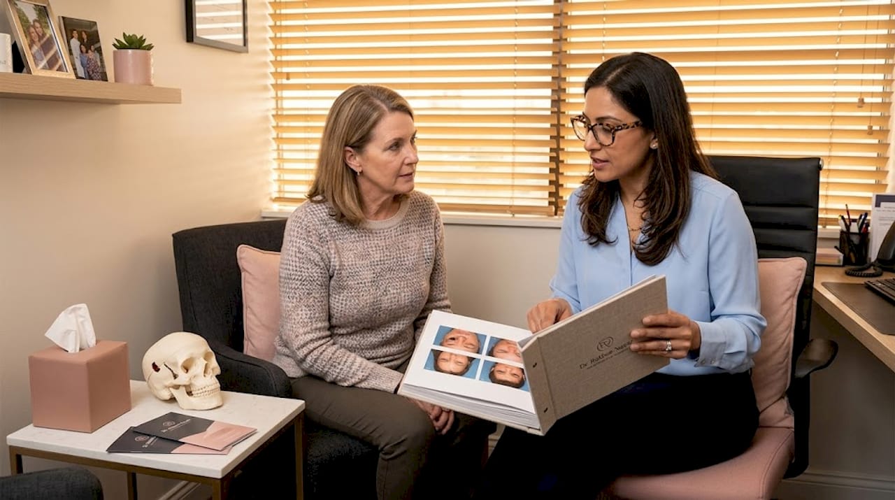
Scarring is a natural part of the healing process, but it can often be optimised. Learn more about minimising scarring after surgery and about restoring confidence after skin cancer through expert reconstruction.
Aftercare and monitoring: what happens next?
Once your wound has been closed and your recovery begins, understanding aftercare means you will heal better and feel more reassured at every stage. Most patients are given detailed written aftercare instructions before leaving the clinic and have access to support through follow-up appointments and their local care team.
The key aftercare essentials to follow include:
- Keep the wound clean and dry for the first 24 to 48 hours as instructed, then follow specific cleaning guidance given by your team
- Watch for signs of infection including increasing redness, swelling, warmth, discharge, or fever, and contact your team promptly if any appear
- Avoid strenuous activity for one to two weeks to reduce the risk of bleeding or wound dehiscence
- Protect the area from sun exposure using high-factor SPF and physical barriers once the wound has healed, as scars are particularly vulnerable to UV damage
- Attend follow-up appointments as scheduled, typically for stitch removal at seven to fourteen days and a wound check shortly after
- Continue skin monitoring and report any new or changing lesions to your GP or specialist
Your care team should give you a clear point of contact in case concerns arise between appointments. Do not hesitate to reach out if anything worries you. For detailed guidance on looking after your skin during recovery, the resource on skin health during Mohs recovery covers practical, stage-by-stage advice.
A surgeon’s perspective: what really matters beyond the surgical steps
Step-by-step guides serve a genuinely important purpose. They reduce anxiety, set expectations, and allow patients to feel like informed participants rather than passive recipients of care. But after years working in Mohs and facial reconstruction, I would be doing patients a disservice if I suggested the steps alone tell the whole story.
Every reconstruction is personal. Two patients with identically sized BCCs in the same location can have quite different conversations about what “the best outcome” means to them. One may prioritise the fastest possible recovery; another may be willing to tolerate a longer healing process for an outcome that is less visible. There is no universal correct answer. The surgeon’s role is to understand what matters to you, not to apply a formula.
This is also why I would encourage every patient to read about Mohs myths and facts before their first consultation. Some assumptions patients carry into clinic, such as the belief that Mohs is always the most invasive option, or that scarring is unavoidable and severe, can create unnecessary worry that gets in the way of honest dialogue.
Ask every question you have. Write them down beforehand if that helps. No reputable surgeon will be irritated by a thorough patient. Informed consent is not a box to tick; it is a foundation for trust, and trust genuinely affects outcomes. Patients who understand what is happening heal with greater confidence and tend to follow aftercare instructions more carefully, both of which matter clinically.
The emotional dimension of facial surgery after a skin cancer diagnosis should never be minimised. It is entirely reasonable to feel anxious, to grieve the change to your skin’s appearance, or to need time to process the experience. Surgical excellence includes recognising and responding to this, not just technical precision at the operating table.
Consult an expert for best results
If you have recently been diagnosed with BCC or have been referred for Mohs surgery, speaking with a specialist who holds dual expertise in both Mohs micrographic surgery and plastic and reconstructive surgery makes a significant difference to your care and your confidence.

Miss Rakhee Nayar offers precisely this combination, with extensive experience in Mohs surgery expertise and facial reconstruction at her clinic in North West England. Whether you want to find out about Mohs before booking a consultation, or are ready to discuss reconstruction after basal cell removal, her team offers in-person and e-consultation options for patients across the UK and internationally. Expert, patient-centred care is the right foundation for the best possible result.
Frequently asked questions
How long does Mohs surgery take for basal cell removal?
Each stage takes approximately 30 minutes for the excision plus up to two hours for laboratory analysis, and some patients are clear in a single stage while others require additional passes, meaning the full day should be kept free.
Will Mohs surgery leave a scar?
Some scarring is expected, but the reconstruction methods available including primary closure, local flaps, and skin grafts, are chosen to minimise visibility and are tailored specifically to your anatomy and preferences.
Is Mohs always necessary for basal cell carcinoma?
Mohs is most appropriate for BCC on the head and neck, recurrent or aggressive subtypes, lesions with unclear borders, and high-risk anatomical locations such as the nose, eyes, and lips.
Can reconstruction be done on the same day as Mohs surgery?
In most cases, yes. Reconstruction is typically performed immediately after clear margins are confirmed, with the chosen method discussed and agreed with the patient before or during the same visit.
What should I do if I have concerns after surgery?
Contact your care team or local clinic immediately if you notice any signs of infection, unexpected pain, or unusual changes to your wound, as prompt review ensures problems are caught and managed early.
Recommended
- How to prepare for Mohs surgery: your step-by-step guide
- Basal cell carcinoma: Mohs surgery and facial aesthetics
- Why choose Mohs surgery for facial skin cancer removal?
- Mohs surgery myths vs facts: what patients need to know
Filed under Mohs Surgery