TL;DR:
- A melanoma diagnosis prompts urgent referral, with biopsy confirming the diagnosis and determining depth. Surgery, including Mohs micrographic procedures for sensitive areas, offers effective treatment with careful reconstruction planning. Systemic therapies enable management of advanced melanoma, while side effect monitoring ensures treatment success and preserves quality of life.
A melanoma diagnosis changes everything in an instant. You’re suddenly navigating referral letters, specialist appointments, and a vocabulary full of terms you’ve never needed before. Melanoma cancer treatment in the UK has advanced considerably, and the options available to you now, from precise surgical removal to systemic immunotherapy, are genuinely effective when applied at the right stage and by the right hands. This guide walks you through the diagnosis pathway, surgical and non-surgical treatments, and the specific role of Mohs micrographic surgery for melanomas in cosmetically sensitive areas, so you can approach your care with clarity rather than anxiety.
Table of Contents
- Understanding melanoma diagnosis and referral
- Standard surgical treatment options for melanoma
- When and how Mohs surgery is used for melanoma treatment
- Alternative and adjunct treatments for melanoma
- Managing side effects and ensuring continued treatment success
- What we have learnt from treating melanoma patients in cosmetically sensitive areas
- Expert melanoma care with Mohs surgery at mohssurgeon.co.uk
- Frequently asked questions
Key Takeaways
| Point | Details |
|---|---|
| Structured referral pathways | UK melanoma referrals are guided by checklist scores and dermoscopy findings, not just visual assessment. |
| Surgical removal basics | Melanoma is primarily treated by excision with safe margins, with surgery tailored to the lesion’s depth and location. |
| Mohs surgery selectivity | Mohs surgery is chosen mainly for melanomas in delicate areas to maximise tissue preservation and clear margins. |
| Non-surgical alternatives | Topical treatments like imiquimod and cryosurgery support cases where surgery is unsuitable or declined. |
| Immunotherapy toxicity management | UK guidelines help manage skin side effects of immunotherapy to keep treatment effective and continuous. |
Understanding melanoma diagnosis and referral
Before any treatment begins, melanoma must be correctly identified and referred through the appropriate NHS pathway. This matters more than most patients realise, because delays at the referral stage directly affect outcomes.
In the UK, your GP does not simply refer based on how a mole looks to the naked eye. Urgent referral is triggered by a weighted 7-point checklist score of 3 or more, or by dermoscopy findings that suggest melanoma. Dermoscopy is a specialised handheld tool that illuminates skin structures invisible to the naked eye. A lesion that looks unremarkable in a standard consultation may score highly under dermoscopy, and vice versa. This is why seeing a GP or dermatologist with access to dermoscopy is so important if you have a changing or suspicious mole.
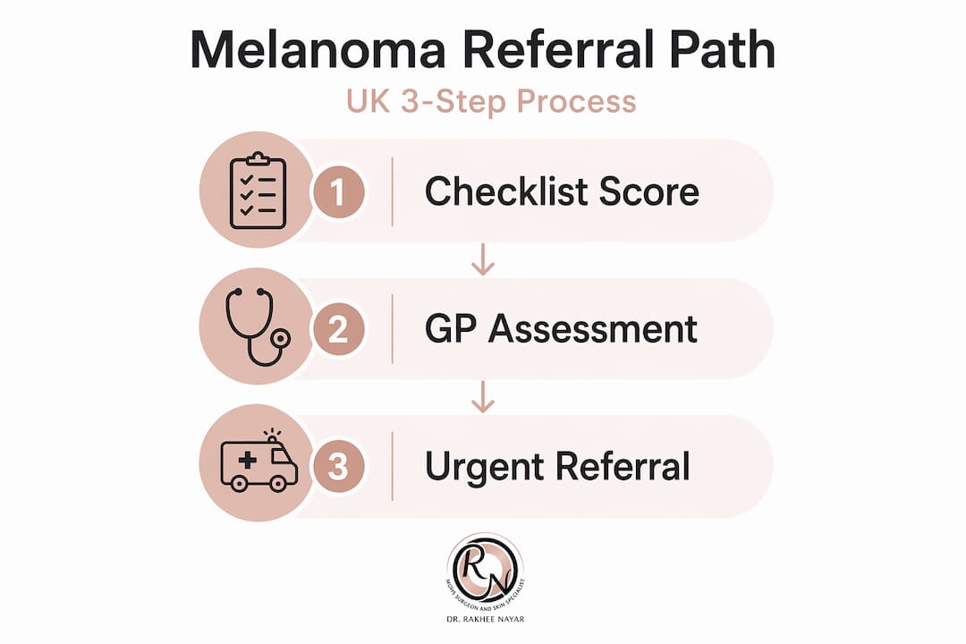
Once referred, diagnosis is confirmed through microscopic examination of tissue taken during an initial narrow-margin excision. This is not the definitive treatment. It is a diagnostic step. A pathologist examines the removed tissue to confirm whether melanoma is present, what subtype it is, and how deeply it has grown into the skin, measured in millimetres as the Breslow thickness.
Key features that may trigger urgent referral include:
- A mole that has recently changed in size, shape, or colour
- Irregular or asymmetric borders
- Multiple shades of brown, black, red, or white within one lesion
- Bleeding, crusting, or ulceration without obvious cause
- A new lesion appearing in someone over 40, particularly on sun-exposed skin
Early skin cancer detection is the single most powerful factor in improving survival outcomes. If you are uncertain about a lesion, do not wait for your next routine appointment.
Standard surgical treatment options for melanoma
With diagnosis confirmed, the next step involves understanding standard surgical options for melanoma treatment. Surgery remains the primary and most effective treatment for the vast majority of melanoma cases, particularly those caught at an early stage.
The standard approach is a wide local excision, which removes the melanoma along with a surrounding rim of healthy skin. The width of that margin depends on the Breslow thickness of the tumour. A melanoma less than 1 mm deep typically requires a 1 cm margin; deeper tumours may need 2 cm or more. This margin is not arbitrary. It exists because melanoma cells can extend microscopically beyond the visible edge of the lesion.
Surgical removal is usually curative for early-stage melanoma when performed promptly and completely. The critical word is “completely.” Incomplete excision, where tumour cells remain at the margins, significantly increases recurrence risk and may require further surgery.
What determines the surgical plan:
- Breslow thickness: The depth of invasion, measured in millimetres, is the most important prognostic factor
- Ulceration: Whether the surface of the tumour has broken down affects staging and margin decisions
- Sentinel lymph node biopsy: For melanomas over 0.8 mm thick, a sentinel node biopsy may be offered to check whether cancer has reached the lymphatic system
- Location: Melanomas on the face, hands, or feet require careful planning to balance clearance with function and appearance
Surgical excision for skin cancer on cosmetically sensitive areas demands a surgeon who can plan reconstruction at the same time as achieving clear margins. This is where specialist training makes a measurable difference.
Pro Tip: Ask your surgeon specifically whether your excision margins will be checked intraoperatively or only after the procedure. Standard excision sends tissue to pathology after surgery, meaning you may need a second operation if margins are not clear. Understanding this upfront helps you plan.
When and how Mohs surgery is used for melanoma treatment
Among surgical options, Mohs micrographic surgery occupies a specific and important place in melanoma care. It is not the default treatment, but for the right patient and the right melanoma, it offers something standard excision cannot: real-time, complete margin assessment during the procedure itself.
Mohs surgery is used for melanoma particularly for lentigo maligna, a slow-growing subtype that tends to appear on sun-damaged facial skin in older adults, and for melanomas in locations where tissue preservation is clinically important. The face, ears, nose, eyelids, and scalp are the most common sites where Mohs becomes the preferred option.

The key difference from standard excision is the margin examination process. In Mohs surgery, thin layers of tissue are removed and examined under a microscope while you wait. If cancer cells are present at any edge, only that specific area is removed in the next layer. This continues until all margins are clear. The result is complete tumour removal with the smallest possible wound, which directly improves the cosmetic and functional outcome of reconstruction.
Comparison of Mohs surgery versus standard wide local excision for melanoma:
| Feature | Mohs micrographic surgery | Standard wide local excision |
|---|---|---|
| Margin assessment | Complete, intraoperative | Partial, post-operative |
| Tissue preservation | Maximum | Fixed margin removed regardless |
| Best suited for | Face, sensitive areas, lentigo maligna | Body, trunk, limbs |
| Reconstruction planning | Immediate, same session | May be staged |
| Risk of second operation | Lower | Higher if margins involved |
Situations where Mohs may be considered for melanoma:
- Lentigo maligna or lentigo maligna melanoma on the face
- Melanoma near the eye, nose, ear, or lip where tissue loss has functional consequences
- Recurrent melanoma where previous scarring makes margin assessment complex
- Patients where cosmetic outcome is a significant clinical consideration
Mohs eligibility for melanoma depends on the subtype and location, so not every melanoma patient will be a candidate. A specialist assessment is essential before assuming Mohs is appropriate for your case. Understanding Mohs versus standard excision in detail helps you ask the right questions at your consultation.
Pro Tip: If your melanoma is on your face and you have been offered standard excision with wide margins, it is entirely reasonable to ask whether Mohs micrographic surgery is appropriate for your case. A second opinion from a Mohs-trained surgeon costs nothing in terms of time lost and may significantly change your outcome.
Alternative and adjunct treatments for melanoma
In cases where surgery is not feasible or melanoma has spread, other treatment options come into play. These range from topical agents for very superficial disease to systemic therapies for advanced or metastatic melanoma.
For melanoma in situ, which is the earliest stage where cancer cells have not yet invaded deeper skin layers, non-surgical options such as imiquimod cream or cryosurgery may be considered when surgery is declined or not possible. Imiquimod is a topical cream that stimulates the immune system to attack cancer cells. It is not a first-line treatment, but it has a role in specific circumstances, particularly for large lentigo maligna on the face where surgery would cause significant disfigurement.
For advanced or metastatic melanoma, systemic therapies have transformed what was once a very poor prognosis into a condition where long-term survival is achievable for a meaningful proportion of patients. The main categories are:
- Immunotherapy: Checkpoint inhibitors such as pembrolizumab and nivolumab block proteins that cancer cells use to hide from the immune system. These are now standard first-line treatments for advanced melanoma and have produced durable responses in patients who previously had very limited options.
- Targeted therapy: For melanomas with a BRAF gene mutation, which accounts for roughly half of all cases, drugs such as dabrafenib and trametinib can directly block the faulty signalling pathway driving tumour growth.
- Radiation therapy: Used when surgery is not feasible, for brain metastases, or to manage symptoms from tumours in specific locations. It is rarely curative for melanoma but plays an important palliative role.
- Chemotherapy: Now largely superseded by immunotherapy and targeted therapy for most patients, but still occasionally used in specific circumstances.
Systemic skin cancer treatments are typically managed by a medical oncologist within a multidisciplinary team. If you are at stage 3 or stage 4, you should expect to be discussed at a skin cancer multidisciplinary team meeting before any treatment plan is finalised.
Managing side effects and ensuring continued treatment success
If you undergo immunotherapy, understanding toxicity management can help you stay on track with treatment. Immunotherapy works by activating your immune system, and that same activation can cause inflammation in healthy tissues, including the skin.
Skin side effects are among the most common immune-related adverse events. They include rashes, itching, blistering, and in rarer cases, severe reactions affecting mucous membranes. Most are manageable, but they need to be recognised and treated promptly to avoid unnecessary breaks in your cancer treatment.
UK consensus guidelines help clinicians manage these skin toxicities with the aim of reducing treatment interruptions. The guidance prioritises topical treatments such as corticosteroid creams before escalating to oral steroids, because systemic immunosuppression can blunt the anti-tumour effect of immunotherapy.
Signs that warrant prompt escalation to your oncology team:
- A rash covering more than 30% of your body surface area
- Blistering or skin that peels in sheets
- Involvement of the mouth, eyes, or genitals
- Skin symptoms accompanied by fever or joint pain
- Any skin change that appears suddenly and worsens rapidly
“Recognising and grading cutaneous toxicities early allows clinicians to intervene with topical treatments before oral steroids become necessary, preserving the efficacy of immunotherapy while protecting the patient’s quality of life.” — Melanoma Focus UK Consensus Guidelines
Pro Tip: Keep a simple photo diary of any skin changes during immunotherapy. Timestamped photos give your oncology team far more useful information than a verbal description and can speed up the decision about whether to escalate or continue treatment unchanged.
What we have learnt from treating melanoma patients in cosmetically sensitive areas
The conversation about melanoma treatment too often focuses exclusively on clearance rates, as though removing the cancer is the only goal. In reality, where and how that removal happens matters enormously to the patient sitting in front of you.
We have seen patients who were offered a 2 cm margin excision on the nose or eyelid, told it was “standard,” and only later discovered that Mohs surgery would have achieved the same clearance with a fraction of the tissue loss. The reconstruction that follows a poorly planned excision on the face can be far more complex, more expensive, and more emotionally difficult than the one that follows a Mohs procedure planned from the outset with reconstruction in mind.
The uncomfortable truth is that not every surgeon offering melanoma treatment has dual training in both oncological excision and plastic reconstruction. Those two skill sets are rarely combined, and the gap shows in outcomes. When you are choosing where to have a melanoma removed from your face, asking about reconstruction planning is not vanity. It is clinical sense.
Expert melanoma care with Mohs surgery at mohssurgeon.co.uk
If you have been diagnosed with melanoma or have a suspicious lesion on a cosmetically sensitive area, specialist assessment can make a real difference to both your cancer outcome and your recovery.

Miss Rakhee Nayar holds dual training in Mohs micrographic surgery and plastic reconstructive surgery, a combination that is rare in the UK and directly relevant to melanoma patients who need both complete tumour clearance and thoughtful reconstruction. Whether you need a Mohs surgery assessment or a second opinion on a planned excision, consultations are available privately at her North West England clinic and via e-consultation for patients across the UK. You do not have to accept a treatment plan that does not fully address both the cancer and the cosmetic consequences.
Frequently asked questions
What triggers urgent referral for suspected melanoma in the UK?
Urgent referral is recommended if a pigmented lesion scores 3 or more on the weighted 7-point checklist or if dermoscopy findings suggest melanoma, regardless of how the lesion appears to the naked eye.
Is Mohs surgery suitable for all melanoma cases?
Mohs is used selectively for melanomas in cosmetically or functionally sensitive areas and for certain subtypes such as lentigo maligna, so suitability must be assessed individually by a specialist with Mohs training.
What are alternatives if surgery is not possible for melanoma in situ?
Imiquimod cream and cryosurgery are recognised alternatives for very superficial melanoma in situ when surgery is declined or contraindicated, though they are not considered equivalent to surgical excision in terms of certainty of clearance.
How are skin side effects from immunotherapy managed in the UK?
Management follows UK consensus guidelines that prioritise topical corticosteroid treatments and early recognition of red-flag symptoms to keep patients on therapy with minimal interruption.