TL;DR:
- Skin cancer mapping includes early detection through mole surveillance and precise removal during Mohs surgery. Mole mapping is non-invasive and monitors high-risk patients over time, while Mohs mapping involves staged excision with microscopic margin control. Both methods improve treatment outcomes, especially on the face, by ensuring complete removal and better cosmetic results.
When people hear “skin cancer mapping,” many assume it simply means checking moles at a routine appointment. In reality, mapping covers two quite different clinical processes, each serving a distinct purpose in your care. One helps detect cancer early through surveillance. The other guides a surgeon’s hands during precise tumour removal. Understanding the difference can genuinely change how you approach a diagnosis, choose a specialist, and think about your recovery. This article walks you through both types, explains what actually happens at each stage, and helps you work out which applies to your situation.
Key Takeaways
| Point | Details |
|---|---|
| Two main mapping types | Skin cancer mapping includes both mole surveillance and precision mapping during surgery. |
| Early detection advantage | Full body mole mapping helps find skin cancer sooner for better outcomes. |
| Surgical accuracy matters | Mohs margin mapping ensures full cancer removal while saving healthy tissue. |
| Facial care priorities | Precise mapping in Mohs surgery maximises both cure and cosmetic results, especially for the face. |
What is skin cancer mapping?
The term “skin cancer mapping” gets used loosely, which causes real confusion for patients trying to research their options. Clinically, it refers to two distinct concepts: full body mole mapping for surveillance of high-risk patients, and tumour margin mapping used during Mohs micrographic surgery to precisely identify and remove remaining cancer cells while sparing healthy tissue.
These two approaches serve completely different purposes, and patients typically encounter them at different points in their journey.
Full body mole mapping is a surveillance tool. It uses total body photography combined with automated lesion tracking to monitor changes in moles over time. It is used before cancer is confirmed, as a way of catching problems early in people who are at elevated risk.
Tumour margin mapping is a surgical tool. It is used once cancer has already been diagnosed and a decision has been made to operate. The surgeon uses colour-coded diagrams and microscopic tissue analysis to track exactly where cancer cells remain in the skin, layer by layer.
Here is a simple comparison of the two approaches:
| Feature | Full body mole mapping | Mohs margin mapping |
|---|---|---|
| Purpose | Early detection and surveillance | Precise surgical removal |
| When used | Before confirmed diagnosis | During confirmed cancer surgery |
| Technique | Photography and digital comparison | Tissue excision, colour-coding, microscopy |
| Who performs it | Dermatologist or trained technician | Mohs surgeon |
| Invasive? | No | Yes, under local anaesthetic |
The key points to remember are:
- Mole mapping does not replace a professional skin examination
- Mohs margin mapping is not used for every skin cancer, only specific types and locations
- Both processes are complementary and may both be relevant to your care at different times
- Neither is exclusively for advanced cases; early-stage patients benefit from both
Understanding the skin cancer detection methods available to you helps you ask better questions at your consultation and make more confident decisions about next steps.
How full body mole mapping works
Full body mole mapping uses total body photography and automated lesion mapping for ongoing surveillance of high-risk patients, with early detection as the primary goal. It is not a one-off event but a longitudinal process, meaning its real value builds over time through comparison between sessions.
Here is how a typical full body mole mapping session works:
- Initial photography session. A trained technician photographs your entire skin surface using standardised lighting and camera positions. This creates a baseline record of every visible lesion, mole, and mark on your body.
- Digital mapping and cataloguing. Software assigns each lesion a reference point and logs its size, shape, colour, and location. This creates a precise digital map unique to you.
- Dermoscopic imaging. Suspicious or high-priority lesions receive close-up dermoscopy images, which capture detail invisible to the naked eye.
- Dermatologist review. A dermatologist reviews the images, flags anything concerning, and compares them against your previous session if one exists.
- Follow-up and monitoring. At your next session, the software compares new images against the baseline. Any lesion that has changed in size, shape, or colour is flagged for clinical review and possible biopsy.
Who benefits most from full body mole mapping? Patients who have a personal or family history of melanoma, those with more than 50 moles, people with fair skin who have had significant sun exposure, and anyone who has previously had a skin cancer removed. It is also valuable for patients who find it difficult to monitor their own skin, such as those with lesions on the back or scalp.
The benefits are meaningful. Early detection dramatically improves treatment outcomes. Mole mapping also reduces unnecessary surgical excisions because decisions are based on documented change rather than clinical suspicion alone. A mole that looks slightly unusual but has been stable for two years is very different from one that has doubled in size over six months.
Pro Tip: Between mapping sessions, photograph any mole that concerns you using your phone and note the date. This gives your dermatologist useful additional context, even if it is not part of a formal mapping programme.
One important limitation: mole mapping is a surveillance tool, not a diagnostic one. It cannot tell you definitively whether a lesion is cancerous. That still requires a biopsy and histological analysis. Think of mapping as an early warning system, not a final answer.
Mohs margin mapping: what happens during surgery
Once skin cancer is confirmed and surgery is planned, a completely different kind of mapping begins. Mohs micrographic surgery uses a precise, staged mapping process that gives surgeons something conventional excision simply cannot offer: the ability to examine 100% of the surgical margins before closing the wound.
In conventional excision, tissue is removed and sent to a laboratory, where pathologists examine a small sample of the margins using a technique called bread-loaf sectioning. This method only examines roughly 1% of the true margin. Mohs mapping, by contrast, examines the entire margin at each stage.
Here is how the steps in Mohs margin mapping work in practice:
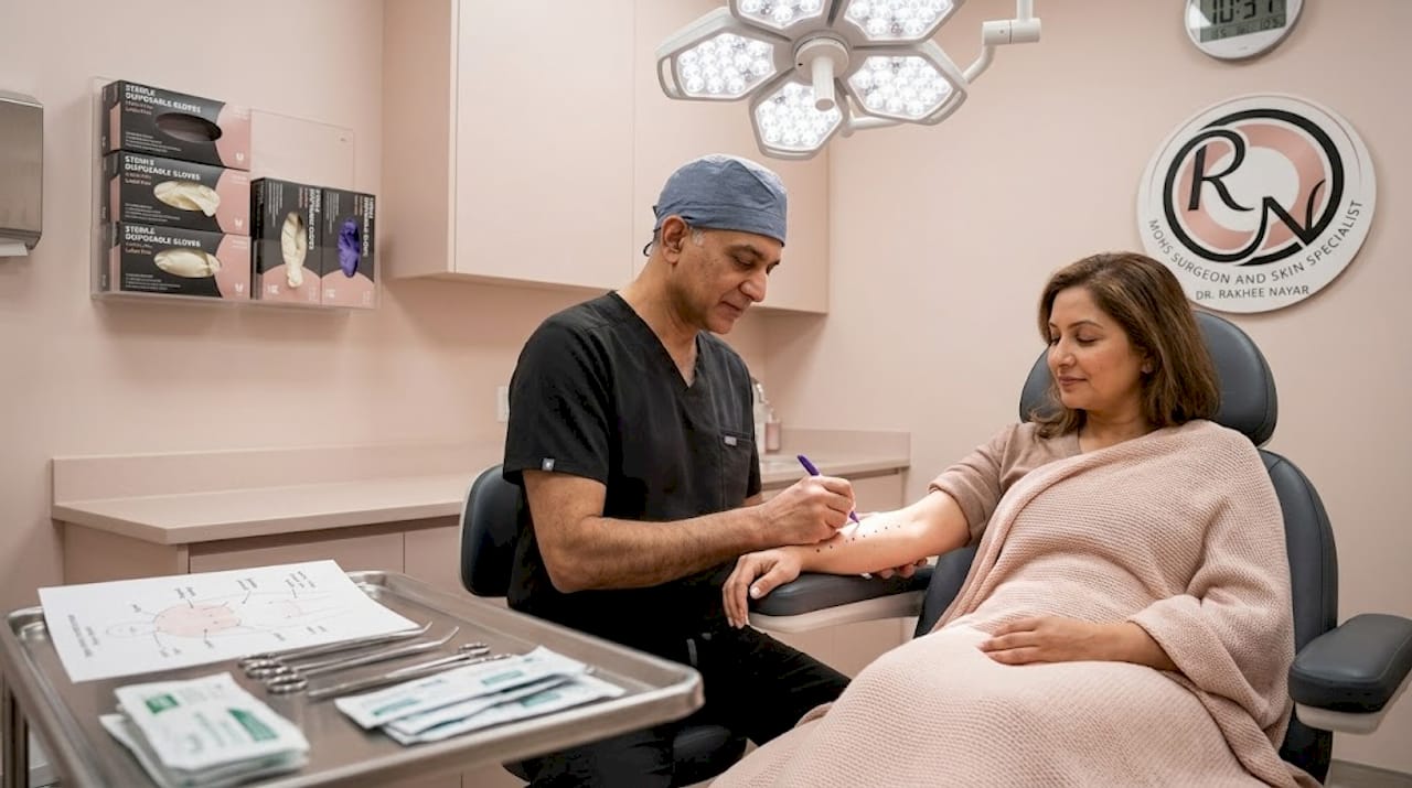
- Visible tumour excision. The surgeon removes the visible tumour with a minimal surrounding margin, cutting at a 45-degree bevel to allow the tissue edges to lie flat for microscopic examination.
- Tissue orientation and division. The excised tissue is carefully oriented using reference marks, then divided into sections. Each section is labelled and colour-coded with dyes to preserve its exact position relative to the patient’s skin.
- 2D mapping diagram. A detailed diagram is drawn, showing exactly where each piece of tissue came from on the patient’s body. This is the “map” itself.
- Frozen section processing. The tissue sections are frozen, cut into thin slices, and mounted on slides. The Mohs surgeon examines these under a microscope, looking for any remaining cancer cells.
- Targeted re-excision. If cancer cells are found, their precise location is marked on the diagram and the surgeon returns to that exact spot on the patient to remove another thin layer. This is repeated until all margins are clear.
“Mohs mapping allows 100% margin control versus bread-loaf sectioning at approximately 1%, making it critical for the Mohs-reconstructive combination in facial cases to optimise both cosmesis and function.” NCBI Mohs surgery essentials
Most cases require between one and three stages to achieve clear margins, though complex tumours may need more. Throughout the procedure, you wait in a comfortable area between stages. The entire process is performed under local anaesthetic.
Pro Tip: Ask your surgeon to explain the mapping diagram before your procedure begins. Seeing the 2D map helps you understand exactly what is being tracked and why the staged approach takes the time it does.
The Mohs surgery explained process is particularly well suited to cancers on the face, ears, nose, and eyelids, where preserving healthy tissue is as important as removing the cancer.
Benefits of skin cancer mapping for surgical and cosmetic outcomes
Precise mapping changes outcomes in ways that matter enormously to patients, particularly when the cancer is on the face or another visible area. The advantages go well beyond simply removing more cancer.
The core benefit of Mohs margin mapping is 100% margin control compared to conventional excision, which is critical for the Mohs-reconstructive combination in facial cases to optimise both cosmesis and function. This is not a minor technical distinction. It means the surgeon knows with certainty that all cancer has been removed before reconstruction begins, rather than discovering positive margins after the wound has already been closed.
Key benefits for patients include:
- Higher cure rates. Mohs surgery achieves cure rates of up to 99% for primary basal cell carcinoma, significantly higher than standard excision.
- Maximum tissue preservation. Because only confirmed cancerous tissue is removed at each stage, healthy skin is spared. This is especially important on the nose, eyelids, lips, and ears where tissue is limited.
- Better reconstructive options. When less healthy tissue is removed, the role of mapping in reconstruction becomes clear. Surgeons have more options for closing the wound naturally, using local flaps or grafts with better cosmetic results.
- Fewer repeat surgeries. Clear margins confirmed intraoperatively mean far fewer cases where patients need a second operation to remove residual cancer.
- Reduced anxiety. Knowing that margins have been thoroughly checked during surgery, rather than waiting days for laboratory results, gives patients significant peace of mind.
Artificial intelligence tools are increasingly used in mole mapping to flag suspicious lesions, and they genuinely improve detection rates. However, AI enhances rather than replaces expert dermoscopy. A skilled specialist brings clinical context, patient history, and tactile assessment that no algorithm currently replicates. The aesthetic considerations after Mohs are best managed by a surgeon with dual training in both oncology and plastic surgery, precisely because the decisions made during mapping directly influence what reconstruction is possible afterwards.
Comparing mole mapping and Mohs mapping: which is right for you?
Understanding which type of mapping applies to your situation helps you have far more productive conversations with your care team. The two processes are not competing options; they serve different moments in your journey. But patients often arrive at consultations unsure which they need, or whether mapping is even relevant to them.
Two distinct concepts define skin cancer mapping in clinical practice: full body mole mapping for surveillance and tumour margin mapping in Mohs surgery. Here is a direct comparison to help you identify where you currently sit:
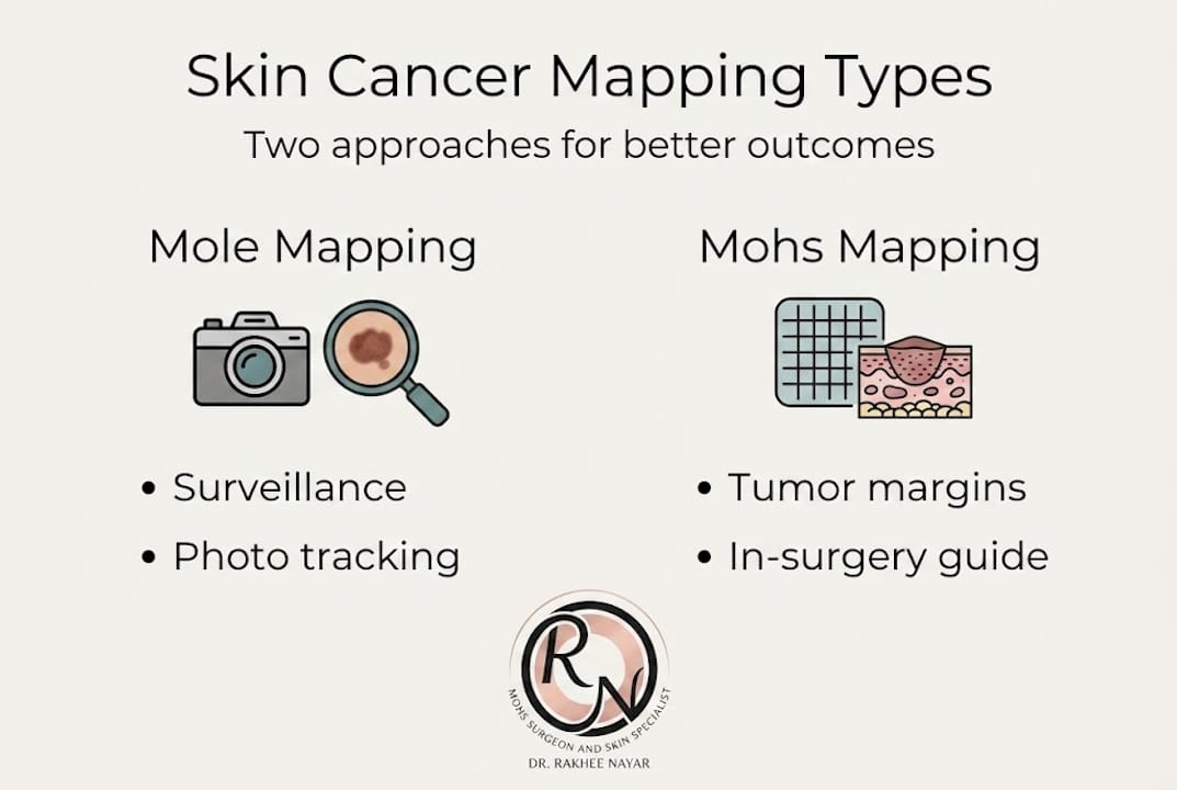
| Full body mole mapping | Mohs margin mapping | |
|---|---|---|
| Your situation | At-risk, no confirmed cancer | Confirmed skin cancer diagnosis |
| Primary goal | Detect changes early | Remove cancer completely |
| Main steps | Photography, comparison, review | Excision, mapping, microscopy, re-excision |
| Outcome | Monitoring plan or referral | Clear surgical margins |
| Specialist needed | Dermatologist | Mohs surgeon |
A common myth is that mapping is only relevant once cancer has become advanced or difficult to treat. In fact, both types of mapping are most effective when used early. Mole mapping catches cancers at their most treatable stage. Mohs mapping is often recommended for early-stage but high-risk tumours in sensitive locations, not just large or recurrent ones.
Questions worth discussing with your specialist include:
- Am I considered high-risk for melanoma or other skin cancers?
- Would full body mole mapping be appropriate for my surveillance?
- If surgery is needed, is my tumour’s location suitable for Mohs?
- What reconstruction options would be available after Mohs in my specific case?
If you have been told you need surgery for a facial skin cancer, understanding Mohs surgery for facial cancer will help you weigh your options clearly. And if you have heard conflicting things about what Mohs involves, reviewing Mohs surgery myths vs facts is a useful starting point before your consultation.
Our perspective: what most guides miss about skin cancer mapping
Most articles about skin cancer mapping focus on the technical mechanics. They explain the photography, the colour-coding, the frozen sections. What they rarely address is why the combination of precise mapping and skilled reconstruction changes not just survival but quality of life.
When we consider advanced facial treatments for skin cancer, the mapping process is not simply a step in the surgical checklist. It is the foundation on which every reconstructive decision rests. A surgeon who understands both the oncological and aesthetic dimensions of mapping can plan reconstruction while still in theatre, making choices that would not be possible if margins were uncertain.
The 100% margin control that Mohs mapping provides is critical for the Mohs-reconstructive combination in facial cases, precisely because it allows the surgeon to close with confidence rather than caution. That confidence translates directly into better cosmetic results and fewer complications.
AI-assisted mole mapping is genuinely useful, but it works best when paired with experienced clinical judgement. The same principle applies in theatre. Technology supports the surgeon; it does not replace the expertise needed to interpret what the map reveals. For patients facing facial reconstruction options, that expertise is what makes the difference between a good outcome and an exceptional one.
Considering Mohs surgery or mapping? Expert care is available
If this article has clarified what skin cancer mapping involves and you are now wondering about your own next steps, specialist care is closer than you might think.

Miss Rakhee Nayar holds dual training in both Mohs surgery and plastic surgery, a combination that is rare in the UK and directly relevant to achieving the best possible outcomes for facial skin cancers. Whether you are seeking surveillance advice, a second opinion on a diagnosis, or planning the Mohs surgery process itself, her clinic in North West England offers private consultations and e-consultations for UK and international patients. For those who need it, facial reconstruction explained is available as part of an integrated treatment plan, ensuring that cancer removal and cosmetic outcome are planned together from the start.
Frequently asked questions
Is skin cancer mapping painful?
Mole mapping is entirely non-invasive and pain-free, while Mohs margin mapping is part of a surgical procedure carried out under local anaesthetic, so discomfort is minimal and well managed.
How often should I have full body mole mapping?
High-risk individuals typically benefit from annual mole mapping, though your dermatologist may recommend a more frequent or tailored schedule based on your personal risk profile.
Is every skin cancer patient offered Mohs margin mapping?
Mohs mapping is recommended for specific cancer types in high-risk or cosmetically sensitive locations; a tissue is oriented and mapped approach is not applied to every skin cancer case.
Why is mapping so important for facial skin cancers?
Mapping enables complete margin control while preserving healthy tissue, which is essential for achieving good cosmetic and functional results when operating on the face.
What if changes are found during mole mapping?
Any flagged changes are reviewed by a dermatologist who can recommend a biopsy or closer monitoring; automated lesion mapping ensures that even subtle shifts between sessions are documented and acted upon appropriately.