
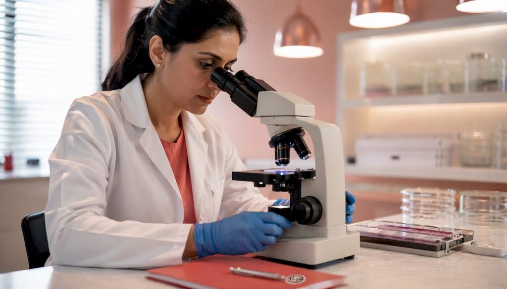
Histopathology is defined as the microscopic examination of tissue to identify disease at a cellular level. In skin cancer, it is the gold standard for definitive diagnosis, providing information that no clinical examination or imaging can replicate: cancer type, Breslow depth, cellular differentiation, and surgical margin status. These findings directly determine whether a patient needs further surgery, sentinel lymph node biopsy, or systemic treatment. The role of histopathology in skin cancer extends from the moment a lesion is removed to every subsequent treatment decision. Miss Rakhee Nayar, GMC-registered Consultant Plastic Surgeon and Mohs surgeon at Circle Cheshire, integrates histopathological analysis into every stage of her clinical practice.
How does histopathology testing work after skin lesion removal?
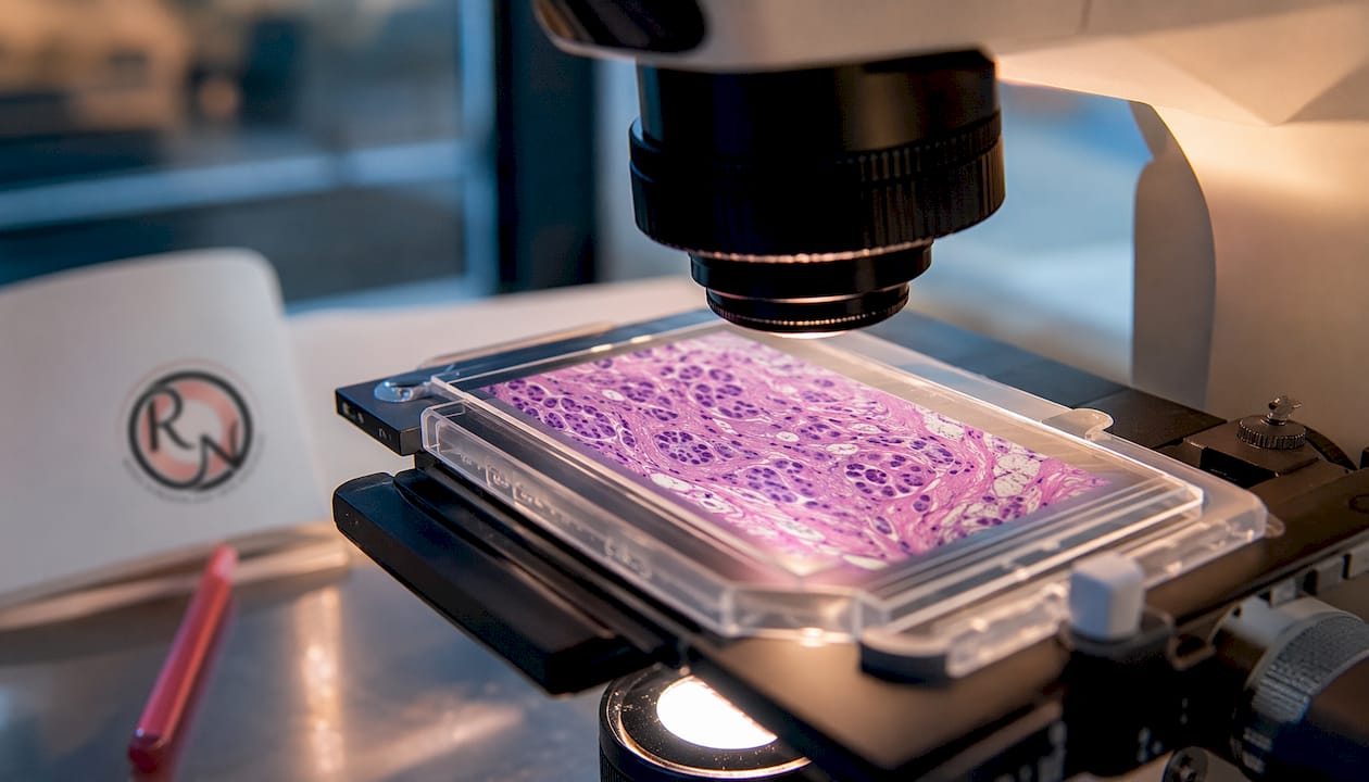
Histopathology begins the moment a tissue sample leaves the operating theatre. The specimen is fixed in formalin, processed into thin sections, mounted on glass slides, and stained using haematoxylin and eosin (H&E). A consultant histopathologist or dermatopathologist then examines the slides under a microscope, assessing cell architecture, nuclear features, invasion depth, and margin status before generating a formal report.
The process follows a defined sequence:
- Specimen fixation. The excised tissue is placed in formalin immediately after removal. Delayed fixation degrades tissue architecture and can compromise diagnostic accuracy.
- Tissue processing and embedding. The fixed specimen is dehydrated, embedded in paraffin wax, and sectioned into slices typically 4–5 micrometres thick.
- H&E staining. Haematoxylin stains cell nuclei blue-purple; eosin stains cytoplasm and connective tissue pink. This contrast allows the pathologist to identify abnormal cells clearly.
- Microscopic examination. The pathologist assesses tumour type, grade, depth of invasion, and whether cancer cells reach the surgical margins.
- Report generation. The written report translates microscopic findings into clinical language, including staging information and recommendations.
The NHS requires 98% of cancer histopathology diagnoses to be reported within 10 days. That target applies to standard H&E analysis. When molecular or next-generation sequencing (NGS) testing is added, the total turnaround extends considerably, as discussed later in this article.
Pro Tip: Ask your clinical team whether your specimen requires additional immunohistochemical or molecular staining. If it does, your report will take longer than the standard 10-day window, and knowing this in advance reduces unnecessary anxiety.
Precise specimen handling is not a minor administrative detail. Crush artefact from forceps, incomplete fixation, or mislabelling can render a specimen uninterpretable, requiring repeat biopsy and delaying diagnosis. Experienced surgical teams follow strict protocols to prevent these errors.
When is histopathology mandatory for skin lesions?
Histopathology is not optional for high-risk or clinically uncertain skin lesions. Mandatory histology testing safeguards patient safety and meets Care Quality Commission (CQC) standards, preventing under-treatment or missed malignancy even when the operating surgeon is clinically confident about the diagnosis.
UK clinical practice guidance updated in 2026 specifies that histopathological confirmation is required in the following situations:
- Any pigmented lesion meeting ABCDE criteria (asymmetry, border irregularity, colour variation, diameter greater than 6mm, evolving appearance)
- Lesions with clinical features of basal cell carcinoma (BCC) or squamous cell carcinoma (SCC), particularly on the face, ears, or scalp
- Recurrent lesions at a previously treated site
- Lesions where the clinical diagnosis is uncertain after dermoscopic assessment
- Any lesion removed from an immunocompromised patient
- High-risk anatomical sites where incomplete excision carries significant morbidity
The consequences of omitting histopathology are serious. A missed diagnosis of infiltrative BCC, for example, can result in extensive subclinical spread before the patient returns with symptoms. Delayed diagnosis of melanoma at a thicker Breslow depth carries a materially worse prognosis. From a medico-legal perspective, failure to submit tissue for analysis in a high-risk clinical scenario constitutes a departure from accepted standards of care.
Dermoscopy and clinical assessment by a consultant plastic surgeon play a critical role in deciding which lesions require mandatory histological examination, balancing diagnostic accuracy and healthcare resource use. In ‘See and Treat’ pathways, where a lesion is excised at the first appointment, the specimen is always sent for histopathological analysis regardless of clinical confidence. This is not defensive medicine. It is the correct standard of care.
Pro Tip: If a clinician removes a skin lesion and does not mention sending it for histopathology, ask directly. You are entitled to know whether your tissue has been submitted for analysis and when the report is expected.
Histopathology also determines the subtype of a cancer, which changes management. Superficial BCC, for instance, is amenable to topical treatment or photodynamic therapy. Infiltrative or morphoeic BCC requires surgical excision with margin control, and Mohs micrographic surgery is the preferred technique for facial lesions. The distinction between these subtypes is invisible to the naked eye. Only histopathology confirms subtypes affecting management decisions.
What histopathological features guide skin cancer treatment?
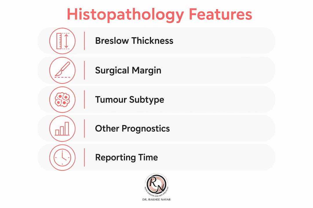
The pathology report is a medical roadmap. It translates microscopic findings into prognosis and treatment information. Patients often find terms like ‘margins’ and ‘Breslow depth’ confusing, yet these are the two most clinically consequential findings in the report.
Breslow thickness
Breslow thickness measures the vertical depth of a melanoma from the top of the epidermis to the deepest tumour cell, expressed in millimetres. It is the single strongest predictor of melanoma prognosis. A melanoma measuring less than 1mm in Breslow depth carries a substantially better prognosis than one measuring 4mm or more. Breslow depth also determines the recommended surgical excision margin and whether sentinel lymph node biopsy is indicated. Patients can understand skin cancer staging by reading the Breslow measurement alongside their T-stage classification.
Surgical margin status
Margin status tells the surgeon whether the cancer was completely removed. A ‘clear’ or ‘negative’ margin means no cancer cells were identified at the cut edge of the specimen. A ‘positive’ or ‘involved’ margin means cancer cells reach the edge, indicating that residual disease may remain in the patient. Understanding surgical margin assessment is central to deciding whether re-excision or Mohs surgery is needed.
Other prognostic features
Several additional histopathological features influence treatment decisions:
- Mitotic rate. The number of cell divisions per square millimetre of tumour tissue. A higher mitotic rate indicates more aggressive tumour biology and upgrades melanoma staging.
- Ulceration. Histological ulceration (not surface trauma) is an independent adverse prognostic factor in melanoma and automatically upgrades the T-stage.
- Lymphovascular invasion. Cancer cells within lymphatic or blood vessels indicate a higher risk of regional or distant spread.
- Perineural invasion. Cancer cells tracking along nerve sheaths, seen most often in SCC, indicate aggressive behaviour and increase the risk of local recurrence.
- Tumour subtype. For BCC, the distinction between nodular, superficial, infiltrative, and morphoeic subtypes determines whether surgery, topical treatment, or radiotherapy is most appropriate.
The pathology report serves as a medical roadmap, translating microscopic findings into prognosis and treatment information. Patients benefit from a clinician walking through each element of the report rather than reading it in isolation. Personalised explanations improve patient engagement and adherence to treatment plans.
How do histopathology delays affect skin cancer outcomes?
Histopathology turnaround time is one of the most consequential but least discussed variables in the skin cancer diagnostic pathway. The NHS target requires 98% of diagnoses within 10 days, but this applies only to standard H&E reporting. When molecular testing is required, such as next-generation sequencing for melanoma subtyping or genetic mutation analysis, the total turnaround averages 17.5–24.5 days. That extension places significant pressure on NHS compliance with the 28-day Faster Diagnostic Standard.
“Histopathology reporting causes major bottlenecks in the cancer diagnostic pathway. The test-to-report interval has historically been treated as a fixed and unalterable phase in NHS trusts, limiting efforts to reduce delays despite its clinical significance. Viewing this interval as an active and optimisable phase can reduce diagnostic delays and improve cancer care pathways.”
Journal of Clinical Pathology, 2025
The clinical consequences of delay are real. A patient waiting beyond 28 days for a confirmed diagnosis experiences measurable psychological distress. More critically, a delay in confirming positive margins after excision of an SCC on the nose, for example, postpones re-excision and allows potential tumour regrowth. For melanoma, a delay in confirming Breslow depth postpones the sentinel lymph node biopsy discussion, which is time-sensitive.
Teledermatology has reduced the need for full diagnostic biopsies by 26%, with biopsy rates falling from 48% to 22% through improved triage. That reduction eases some pressure on histopathology laboratories. Digital pathology, where whole-slide images are analysed remotely by specialist dermatopathologists, offers further potential to reduce bottlenecks. However, investment in digital infrastructure across NHS trusts remains uneven.
For patients navigating the skin cancer specialist referral pathway, understanding that histopathology turnaround is a distinct phase, separate from the surgical waiting time, helps set realistic expectations. Private consultant-led care typically delivers histopathology results faster, with direct clinician communication at each stage.
Key takeaways
Histopathology is the definitive diagnostic method in skin cancer, providing margin status, Breslow depth, tumour subtype, and prognostic features that directly determine every subsequent treatment decision.
| Point | Details |
|---|---|
| Gold standard diagnosis | Histopathology confirms cancer type, depth, and margins; no clinical examination replaces it. |
| Mandatory for high-risk lesions | UK 2026 guidance requires histopathological confirmation for pigmented, recurrent, and uncertain lesions. |
| Report features drive treatment | Breslow depth, margin status, mitotic rate, and ulceration each alter staging and surgical planning. |
| NHS turnaround targets | Standard H&E reporting targets 10 days; molecular testing extends this to 17.5–24.5 days on average. |
| Delays affect outcomes | Histopathology bottlenecks are the leading cause of 28-day Faster Diagnostic Standard non-compliance. |
Histopathology, Mohs surgery, and your next step
Mohs micrographic surgery is the only surgical technique that combines tumour removal and real-time histopathological analysis in a single procedure. Each layer of tissue removed is immediately processed, stained, and examined under the microscope before the next layer is taken. This means the surgeon knows, within the same appointment, whether the cancer has been completely cleared.

Miss Rakhee Nayar is dual-trained in plastic surgery and Mohs surgery, bringing both oncological precision and reconstructive expertise to every case. For patients with BCC or SCC on the face or other cosmetically sensitive areas, Mohs micrographic surgery offers the highest cure rates available alongside careful attention to cosmetic outcome. Private consultations and e-consultations are available at Circle Cheshire for UK-based and international patients. To discuss your histopathology results or explore your treatment options, contact the clinic directly to arrange an appointment.
This article is for informational purposes only and does not constitute medical advice. Consult a GMC-registered specialist for assessment and treatment recommendations.
FAQ
What is histopathology in skin cancer?
Histopathology is the microscopic examination of skin tissue to identify and characterise cancer cells. It is the definitive method for confirming a skin cancer diagnosis, determining tumour type, depth, and whether surgical margins are clear.
Why is histopathology the gold standard for skin cancer diagnosis?
Clinical examination and dermoscopy can raise suspicion of skin cancer, but only histopathology provides a confirmed diagnosis with full staging information. It reveals features such as Breslow depth, mitotic rate, and subtype that directly guide treatment decisions.
How long does a skin cancer histopathology report take?
The NHS targets reporting of 98% of cancer histopathology results within 10 days. When molecular or next-generation sequencing testing is required, the total turnaround typically extends to 17.5–24.5 days.
What does a positive margin mean on a histopathology report?
A positive margin means cancer cells were identified at the cut edge of the removed tissue, indicating that residual tumour may remain. Re-excision or Mohs surgery is usually recommended to achieve clear margins.
Is histopathology always required after mole or lesion removal?
Histopathology is mandatory for any lesion with features of malignancy, any pigmented lesion meeting ABCDE criteria, recurrent lesions, and clinically uncertain diagnoses. UK CQC standards and 2026 clinical guidance require submission of tissue for analysis in all high-risk scenarios.
Recommended
- How is skin cancer staged: a patient’s guide
- High-risk skin cancer: Expert treatment guide for UK patients
- Understanding skin cancer mapping for precise treatment
- Skin cancer symptoms and signs: the UK guide for early detection
Filed under Mohs Surgery


