Basal cell carcinoma (BCC) is the most common facial skin cancer, accounting for around 80% of cases on the face due to cumulative sun exposure. Squamous cell carcinoma (SCC) is the second most frequent type, particularly in patients over 50. Melanoma and rarer cancers such as Merkel cell carcinoma also occur on the face, though less often. Searching for skin cancer on face pictures can raise your awareness of what to look for, but the British Association of Dermatologists and NICE both confirm that only a biopsy can provide a definitive diagnosis. The ‘Ugly Duckling’ sign, noticing a lesion that simply looks different from your other spots, is often more reliable than memorising complex visual rules. This guide explains what each cancer type looks like, where it tends to appear, and when to seek expert assessment.
1. What does basal cell carcinoma look like on the face?
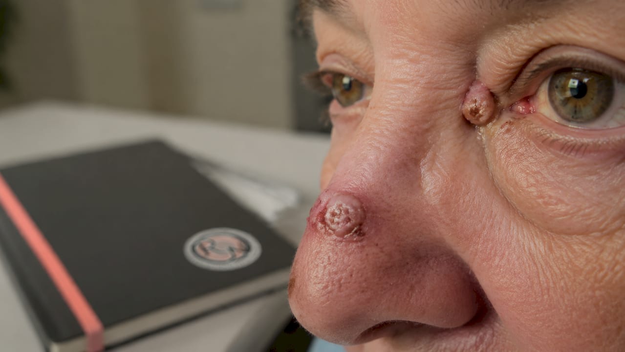
BCC is the most frequently encountered facial skin cancer, and its appearance is often subtler than patients expect. BCC commonly presents as a small, shiny, pearly or translucent pink bump with visible tiny blood vessels called telangiectasias running across its surface. That pearlescent quality is the single most distinctive feature. Many patients initially mistake it for a pimple that simply will not clear.
Common visual presentations of BCC on the face include:
- Pearly or waxy nodule. A small, dome-shaped, shiny bump, often skin-coloured or pale pink, most frequently found on the nose, eyelids, cheeks, forehead, or ears.
- Non-healing sore. A sore that crusts, bleeds, appears to heal, then reopens repeatedly over weeks or months.
- Flat pink or reddish patch. A pale, slightly scaly area that resembles eczema or psoriasis but does not respond to standard topical treatments.
- Scar-like patch. A waxy, pale, ill-defined area that looks like a small scar with no history of injury. This morphoeic subtype is particularly easy to overlook.
- Rolled or raised border. A lesion with a slightly raised, rolled edge around a central depression or ulcer.
The nose, inner corner of the eye, and lower eyelid are especially high-risk sites because they receive intense, reflected sunlight throughout a person’s lifetime. You can view clinical BCC photographs that illustrate these presentations in a UK clinical context.
Pro Tip: If a spot on your face has bled, crusted, and then appeared to settle more than twice in the same location, treat it as suspicious regardless of its size. BCC rarely causes pain, which is precisely why patients delay seeking advice.
2. How to identify squamous cell carcinoma on the face
SCC is the second most common facial skin cancer, and its visual features differ meaningfully from BCC. SCC incidence rises significantly in people over 50, particularly those with a history of outdoor work, sunbed use, or immunosuppression. Unlike BCC, SCC tends to grow more quickly and carries a greater risk of spreading if left untreated.
Typical visual features of SCC on the face include:
- Firm, dome-shaped growth. A raised, reddish nodule with an irregular or rough surface, often appearing on the lower lip, ear, scalp, or cheek.
- Scaly or crusted patch. A persistent, rough, dry area that may bleed when touched or picked. It does not resolve with moisturiser or topical steroids.
- Central ulceration. A crater-like depression in the centre of a raised growth, sometimes described as a wart-like nodule with a hollowed core.
- Cutaneous horn. SCC may present as a horn-like, quickly growing mass or a thick, pigmented, thickened patch, particularly on sun-damaged skin.
- Non-healing sore. A sore that ulcerates, partially heals, and then breaks down again, often over several weeks.
The lower lip is a site that patients frequently underestimate. A persistent roughness or thickening on the lip border, especially in someone who has spent years working outdoors, warrants prompt review. For detailed information on diagnosis and treatment options, the SCC diagnosis and Mohs surgery page provides a thorough clinical overview.
The ABCDE rule, designed primarily for melanoma, is less helpful for SCC and BCC. The ABCDE criteria are less reliable for common facial skin cancers like BCC and SCC, which present as persistent sores or pearly bumps rather than pigmented lesions. Focusing on persistence, change, and failure to heal is more clinically useful for these two cancer types.
3. Recognising melanoma and other rarer skin cancers on the face
Melanoma is less common on the face than BCC or SCC, but it demands urgent attention because of its potential to spread rapidly. Melanoma can appear on the nose, outer ear, eyelid, and less commonly across other facial sites, often in an irregular form that differs from a patient’s other moles.
The ABCDE criteria remain the primary framework for melanoma recognition:
- Asymmetry. One half of the lesion does not match the other in shape or outline.
- Border irregularity. The edges are ragged, notched, blurred, or poorly defined rather than smooth and round.
- Colour variation. The lesion contains more than one shade, including brown, black, red, white, or blue within the same spot.
- Diameter. A lesion wider than 6 millimetres (roughly the size of a pencil eraser) warrants review, though melanomas can be smaller.
- Evolving. Any change in size, shape, colour, or surface, or any new symptom such as bleeding or itching, is a red flag regardless of the lesion’s other features.
Melanoma on the face may appear as a flat or slightly raised irregular mole with uneven colouring. Nodular melanoma is a particularly aggressive subtype that can arise as a rapidly growing, dark or even skin-coloured raised lump, most often on the nose or ear.
Merkel cell carcinoma is a rarer but aggressive facial skin cancer. It typically presents as a fast-growing, firm, flesh-coloured or bluish-red nodule, most often on the face or neck in older patients. It is frequently mistaken for a cyst or lipoma, which is why any rapidly enlarging facial lump in a patient over 60 should be assessed without delay.
If you notice a lesion that is changing, bleeding, or simply looks unlike anything else on your skin, seek a specialist opinion promptly. Do not wait for it to reach a particular size.
4. How to use images effectively to recognise skin cancer on the face
Images of skin cancer on the face serve one clear purpose: they raise awareness and prompt earlier consultation. They do not replace clinical examination. Only a biopsy can confirm a skin cancer diagnosis. Images are for awareness and initial suspicion only. This distinction matters because many benign conditions, including sebaceous cysts, dermatofibromas, and solar keratoses, can closely resemble malignant lesions in photographs.
The most practical use of personal photography is monitoring change over time. Take a clear, well-lit photograph of any lesion you are concerned about and repeat it monthly. If the lesion grows, changes colour, or develops a new texture, you have objective evidence to share with a specialist. That record can significantly shorten the time to diagnosis.
Pro Tip: Use natural daylight rather than a phone flash when photographing a lesion. Flash flattens surface texture and can obscure the pearlescent quality that characterises BCC. A macro setting, if your phone supports it, captures surface detail far more accurately.
“Technological advances including AI-assisted image analysis improve diagnostic support, but they do not replace histopathological confirmation by biopsy, which remains the definitive diagnostic standard for skin cancers on the face.” Current advances in skin cancer diagnosis
Select credible image sources when researching what facial skin cancer looks like. The British Association of Dermatologists, NHS, and Cancer Research UK all publish clinically reviewed photographs. Avoid relying on unmoderated social media images, which may be mislabelled or unrepresentative of typical presentations.
The ‘Ugly Duckling’ sign is often more clinically reliable than memorising complex appearance rules. If one spot on your face simply looks different from all your others, that difference alone is sufficient reason to have it assessed. You do not need to match it precisely to a photograph.
5. Common mistakes when identifying facial skin cancer from pictures
The most frequent error patients make is relying on colour alone. A lesion that is skin-coloured or pale pink does not look dramatic in a photograph, yet BCC and SCC are frequently exactly that. Patients dismiss these lesions because they do not resemble the dark, irregular moles shown in melanoma awareness campaigns.
Common mistakes include:
- Waiting for a lesion to look “serious.” Facial skin cancers often appear as persistent pimples or scar-like areas that patients ignore for months. Most do not look like large tumours or dramatic growths at the stage when treatment is most straightforward.
- Confusing a persistent spot with acne. A pimple that does not resolve within four weeks, particularly in a patient over 40, is not acne until proven otherwise.
- Ignoring a changing lesion because it is small. Size is not a reliable indicator of malignancy. Early-stage SCC and nodular melanoma can both be under a centimetre when they first become clinically significant.
- Assuming a scar-like area is the result of old injury. Morphoeic BCC presents as a pale, waxy, ill-defined patch that patients frequently attribute to a forgotten knock or scratch.
- Treating a non-healing sore with over-the-counter creams. Applying antiseptic or steroid cream to a lesion that repeatedly breaks down delays diagnosis and can temporarily alter its appearance, making clinical assessment harder.
Clinical context matters as much as visual appearance. A GMC-registered specialist considers your age, skin type, sun exposure history, and immune status alongside what they see. No photograph captures that context. The UK guide to skin cancer symptoms sets out the full range of warning signs and explains when each warrants urgent referral.
Key takeaways
Early recognition of facial skin cancer depends on understanding what each type looks like and knowing the limits of visual comparison alone.
| Point | Details |
|---|---|
| BCC is the most common facial cancer | It appears as a pearly, shiny nodule or non-healing sore, most often on the nose, eyelids, and cheeks. |
| SCC grows faster and ulcerates | Look for firm, crusted, dome-shaped growths or persistent rough patches that bleed or fail to heal. |
| Melanoma requires ABCDE assessment | Asymmetry, border irregularity, colour variation, diameter, and evolution are the key warning features. |
| Images raise awareness but cannot diagnose | Only a biopsy confirms skin cancer; use photographs to monitor change and support specialist consultation. |
| The ‘Ugly Duckling’ sign is highly reliable | Any lesion that looks markedly different from your other spots warrants prompt professional assessment. |
What I have learnt from years of treating facial skin cancer
By Miss Rakhee Nayar, GMC-registered Consultant Plastic Surgeon, FRCS (Plast), MD
The patients I see most often are not the ones who ignored an obvious growth. They are the ones who noticed something small, looked it up online, found a photograph that did not quite match, and concluded they were probably fine. That gap between “it doesn’t look like the pictures” and “I should get it checked” is where delayed diagnoses happen.
Facial skin cancers are, by nature, subtle in their early stages. BCC in particular can look almost nothing like the textbook images for months or even years. I have seen lesions dismissed as dry skin, old scars, or persistent spots that turned out to be morphoeic BCC requiring significant reconstruction. The face is not a forgiving site for delayed treatment. The closer a cancer sits to the eye, nose, or lip, the more complex the surgery becomes when it is caught late.
What I encourage every patient to do is take a photograph of anything that concerns them and bring it to a consultation. That image, combined with a clinical examination and, where indicated, a biopsy, gives us the full picture. No AI tool, no online image gallery, and no symptom checker replaces that process. What they can do is give you the confidence to make the appointment.
Modern Mohs micrographic surgery and facial reconstruction mean that even cancers in cosmetically sensitive locations can be treated with high cure rates and good aesthetic outcomes. Early detection makes that significantly easier. If something on your face has been there for more than four weeks and is not resolving, please have it assessed. That is not an overreaction. That is exactly the right response.
— Miss Rakhee Nayar
Specialist assessment for facial skin cancer at Mohs Surgeon UK
Recognising a suspicious lesion is the first step. Getting it assessed by a specialist is the one that matters.

Miss Rakhee Nayar offers private consultations and e-consultations for patients across the UK and internationally, with in-person appointments at her clinic in North West England. Her dual training in plastic surgery and Mohs surgery means that diagnosis, removal, and facial reconstruction are managed by a single expert. Whether you have noticed a persistent sore, a changing mole, or a lesion that simply does not look right, the skin cancer symptoms UK guide provides a clear starting point, and a consultation provides the clinical answer. Private consultations are available; fees are confirmed at the time of booking.
This article is for educational purposes only and does not constitute medical advice. Please consult a GMC-registered specialist for assessment of any skin lesion.
FAQ
What does early skin cancer look like on the face?
Early facial skin cancer most commonly appears as a small, shiny, pearly bump, a non-healing sore, or a persistent rough patch. BCC and SCC rarely look dramatic at an early stage, which is why any lesion that does not resolve within four weeks warrants professional assessment.
Can I diagnose skin cancer from photos alone?
No. Biopsy is the only definitive method for confirming a skin cancer diagnosis. Photographs raise awareness and help you monitor change, but many benign conditions closely mimic skin cancer in images.
Which part of the face is most affected by skin cancer?
The nose, eyelids, ears, forehead, and cheeks are the most commonly affected sites, reflecting cumulative sun exposure across a lifetime. The nose alone accounts for a disproportionately high number of BCC cases.
What is the ABCDE rule and does it apply to all facial skin cancers?
The ABCDE rule covers Asymmetry, Border, Colour, Diameter, and Evolution and is most useful for identifying melanoma. It is less reliable for BCC and SCC, which typically present as persistent sores or pearly nodules rather than pigmented lesions.
When should I see a specialist about a facial lesion?
See a GMC-registered specialist if a lesion has been present for more than four weeks without resolving, bleeds or crusts repeatedly, is changing in size or colour, or simply looks different from your other spots. Early assessment leads to simpler treatment and better outcomes.