
NHS skin cancer images are clinically verified photographs depicting the typical appearances of basal cell carcinoma (BCC), squamous cell carcinoma (SCC), and melanoma, used to support public awareness and early recognition of suspicious lesions. These images form part of NHS skin cancer education materials and are widely referenced by Cancer Research UK, NHS trusts, and specialist dermatology services. Understanding what each cancer type looks like is the first step in knowing when to seek professional advice. This guide presents the key visual features of each cancer type, explains how to use skin cancer photos responsibly for self-monitoring, and outlines when clinical assessment is necessary.
1. What NHS skin cancer images actually show
NHS skin cancer images are not a diagnostic tool. They are educational references that illustrate the range of appearances a given cancer type may present with across different skin tones, body sites, and stages of development. The three cancers most commonly depicted are basal cell carcinoma, squamous cell carcinoma, and melanoma. Each has distinct visual characteristics, though considerable overlap exists with benign lesions such as seborrhoeic keratoses, dermatofibromas, and benign naevi.
The value of these images lies in pattern recognition. When you see a lesion on your own skin and compare it against skin cancer types images, you are not diagnosing yourself. You are deciding whether a lesion warrants professional review. That distinction matters. Overconfidence in image-based self-assessment leads either to unnecessary anxiety or, more dangerously, to false reassurance.
2. Basal cell carcinoma images and their distinctive features
Basal cell carcinoma is the most common skin cancer in the UK and the type most frequently illustrated in NHS skin cancer education materials. BCC typically appears as a pearly or translucent dome-shaped nodule, often with fine blood vessels (telangiectasia) visible across its surface. On darker skin tones, it may appear pigmented and be mistaken for a mole. On fair skin, it often has a waxy, almost skin-coloured sheen.
BCC presents in several distinct subtypes, each with a different visual profile:
- Nodular BCC: The most common subtype. Appears as a raised, round, pearly nodule, sometimes with a central depression or ulcer. Often found on the nose, cheeks, or forehead.
- Superficial BCC: Presents as a flat, scaly, pinkish-red patch with a slightly raised border. Easily confused with eczema or psoriasis.
- Morphoeic (sclerosing) BCC: Appears as a pale, scar-like plaque with ill-defined edges. This subtype is the most difficult to identify visually and is frequently underestimated in size.
- Pigmented BCC: Contains brown, blue, or black pigmentation and can resemble a melanoma or a benign pigmented lesion.
When reviewing skin cancer photos of BCC, pay attention to the border. BCC borders are typically rolled or pearlescent, which distinguishes them from the flat, irregular borders of superficial spreading melanoma. The presence of telangiectasia crossing the surface of a nodule is a particularly reliable visual clue.
Pro Tip: When photographing a suspected BCC, take the image in natural daylight at a 90-degree angle to the skin surface. Place a ruler or coin beside the lesion for scale. This single step significantly improves the usefulness of your photo during a consultation.
3. Squamous cell carcinoma images and how to recognise them visually
Squamous cell carcinoma is the second most common skin cancer in the UK and tends to develop on sun-exposed areas including the face, ears, scalp, and backs of the hands. SCC often appears as a rough, scaly patch or a firm, raised nodule with a crusted or ulcerated surface. Unlike BCC, SCC may bleed spontaneously or fail to heal after minor trauma.
The visual range of SCC is broader than many patients expect:
- Scaly, erythematous patches: These may resemble actinic keratoses (pre-cancerous lesions) and are often found on the lower lip, temples, or dorsum of the hand.
- Firm nodules with central ulceration: These are more advanced presentations and may have a crater-like appearance.
- Cutaneous horn: A hard, horn-like protrusion of keratin arising from a red or inflamed base. This is a recognised SCC variant.
- Pigmented SCC: Less common but can mimic melanoma in appearance.
- Verrucous SCC: A warty, cauliflower-like growth that may be mistaken for a viral wart.
One feature that distinguishes SCC from BCC in skin cancer photos is texture. SCC surfaces are typically rougher and more keratinised, whereas BCC tends to appear smoother and more translucent. Visual assessment alone cannot reliably diagnose skin cancer, as many benign lesions closely mimic malignancies. Professional examination and biopsy remain the diagnostic gold standard.
Any scaly or crusted lesion that has been present for more than four weeks, particularly on a sun-exposed site in a patient over 50, warrants prompt GP referral. The NHS recommends consulting your GP if a skin change persists beyond this timeframe.
4. Melanoma skin cancer images and critical visual warning signs
Melanoma is the most serious of the three common skin cancer types and accounts for the majority of skin cancer deaths in the UK. Melanoma typically shows asymmetry, irregular borders, and varied pigmentation ranging from brown and black through to pinkish or red tones. The ABCDE framework (Asymmetry, Border, Colour, Diameter, Evolution) was developed specifically to make melanoma visual features accessible to the public.
Key visual characteristics seen in melanoma skin cancer images include:
- Asymmetry: One half of the lesion does not mirror the other.
- Irregular borders: Edges are notched, scalloped, or poorly defined rather than smooth and round.
- Colour variation: Multiple shades within a single lesion, including tan, brown, black, red, white, or blue.
- Diameter: Most melanomas are larger than 6mm at diagnosis, though early lesions may be smaller.
- Evolution: Any lesion that has changed in size, shape, colour, or sensation over weeks to months.
Amelanotic melanoma is a particularly challenging variant. It contains little or no pigment and may appear pink, red, or skin-coloured, making it visually indistinguishable from a benign lesion or an inflamed cyst. This subtype is frequently missed on visual inspection alone and reinforces why early skin cancer detection requires professional dermoscopic assessment rather than image comparison alone.
Nodular melanoma, another aggressive subtype, grows rapidly and may appear as a dark, raised nodule without the classic ABCDE features. NHS dermatology resources include images of these atypical presentations precisely because they are the ones most likely to be overlooked.
5. How to use skin cancer images safely for self-monitoring
NHS skin cancer images are educational resources, not diagnostic instruments. Using them correctly means understanding their purpose: to prompt awareness, not to provide a verdict. The following approach reflects current NHS guidance and the recommendations of Cancer Research UK.
Performing a regular skin self-examination
No national screening programme exists for non-melanoma skin cancer in the UK. Self-examination is therefore the primary mechanism by which patients detect new or changing lesions between GP appointments. Perform a full-body check monthly, using a full-length mirror for the front and back of the body and a hand-held mirror for the scalp, behind the ears, and the soles of the feet. Using a hand-held mirror or asking a partner to check difficult areas improves thoroughness, particularly for the back and posterior scalp.
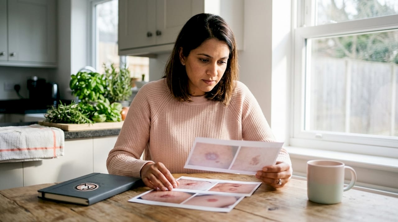
Documenting suspicious lesions with photographs
Patients should photograph suspicious lesions with a ruler or tape measure placed beside the lesion to maintain an accurate size record. These photos allow you to track whether a lesion has grown, changed colour, or developed new features between appointments. A photograph taken in consistent lighting conditions, at the same angle, and at regular intervals creates an objective record that is far more informative than a verbal description.
When to seek medical advice
The following features warrant GP referral without delay:
- A sore or skin change that has not healed within four weeks
- A lesion that bleeds without injury or repeatedly crusts over
- A mole or pigmented lesion that has changed in size, shape, or colour
- A new lesion that looks different from your other moles or spots
- Any lesion causing persistent itch, pain, or altered sensation
What happens after referral
Following GP referral, NHS diagnostic procedures include dermoscopy, biopsy, and imaging. Dermoscopy is a magnified, non-invasive examination that helps clinicians distinguish benign from malignant lesions before any tissue is removed. If dermoscopy raises concern, a biopsy provides definitive tissue diagnosis. CT, MRI, or ultrasound imaging may follow if spread is suspected.
Pro Tip: Create a simple photo log using your smartphone. Label each image with the date, body site, and approximate size. Share this log directly with your GP or specialist at your appointment. Patient-held photographic records improve clinical consultations by providing objective evidence of lesion progression.
Comparison of key visual features by cancer type
| Cancer type | Typical appearance | Key distinguishing feature |
|---|---|---|
| Basal cell carcinoma | Pearly nodule, flat pink patch, scar-like plaque | Rolled, translucent border with telangiectasia |
| Squamous cell carcinoma | Rough scaly patch, crusted nodule, cutaneous horn | Keratinised surface, failure to heal |
| Melanoma | Pigmented lesion with colour variation and irregular border | Asymmetry, multiple colours, rapid evolution |
| Amelanotic melanoma | Pink or red nodule, may resemble a cyst | Absence of pigment despite malignant behaviour |
Key takeaways
Recognising skin cancer visually requires familiarity with the distinct features of BCC, SCC, and melanoma, combined with a structured self-examination routine and prompt referral for any lesion that persists or changes beyond four weeks.
| Point | Details |
|---|---|
| NHS images are educational, not diagnostic | Use skin cancer photos to prompt awareness, not to reach a self-diagnosis. |
| BCC has a pearlescent, rolled border | Telangiectasia crossing the surface is a reliable visual indicator of BCC. |
| SCC surfaces are rough and keratinised | Any scaly, non-healing lesion on a sun-exposed site warrants GP review. |
| Amelanotic melanoma lacks pigment | Pink or red nodules without obvious pigmentation can still be melanoma. |
| Photograph lesions with a ruler | Scaled photos give clinicians objective evidence of size and progression over time. |
Why images alone are never enough: a consultant’s perspective
Patients often arrive at my clinic having spent hours comparing their lesion against NHS skin cancer images online. Some are reassured by what they see. Others are convinced they have a melanoma when the lesion turns out to be a benign seborrhoeic keratosis. Both outcomes concern me, because both reflect a misunderstanding of what clinical photographs are designed to do.
Images are calibrated for pattern recognition across a population. Your lesion exists on your skin, in your context, with your history of sun exposure, previous skin cancers, and immune status. A photograph cannot capture the subtle surface texture that dermoscopy reveals, nor the way a lesion feels under a gloved finger. I have seen BCCs that looked entirely benign in photographs and melanomas that were dismissed for months because they lacked the classic ABCDE features.
What I encourage patients to do is use NHS skin cancer images as a prompt, not a verdict. If something on your skin looks different from everything around it, or if it has changed, that is enough reason to seek an opinion. You do not need to match it precisely to a photograph before you pick up the phone. The photograph is a starting point for a conversation, not the end of one.
I also find that patients who bring a photo log to their first consultation give me something genuinely useful. A series of dated, scaled photographs showing a lesion over three to six months tells me far more than a description. Evaluating skin lesions accurately depends on this kind of objective evidence. If you are monitoring something at home, photograph it consistently and bring those images with you.
— Miss Rakhee Nayar
Concerned about a lesion? Expert assessment at Rakhee Nayar – Mohs Surgeon and Skin Specialist
If a lesion has prompted you to search for NHS skin cancer images, it deserves professional review rather than continued self-monitoring.

Rakhee Nayar – Mohs Surgeon and Skin Specialist offers private consultations and e-consultations at Circle Cheshire in North West England, led by Miss Rakhee Nayar, GMC-registered Consultant Plastic Surgeon, FRCS (Plast), MD. Miss Nayar is dual-trained in plastic surgery and Mohs micrographic surgery, and uses clinical photography as part of her initial triage process. For confirmed BCC or SCC, Mohs micrographic surgery offers precise, margin-controlled removal with same-day results. For patients weighing their options, a detailed comparison of Mohs versus standard excision is available on the website. Private consultation fees are available on request. This article does not constitute medical advice. Please consult a GMC-registered specialist for assessment of any suspicious skin lesion.
FAQ
What do NHS skin cancer images show?
NHS skin cancer images illustrate the typical visual appearances of basal cell carcinoma, squamous cell carcinoma, and melanoma across a range of skin tones and body sites. They are designed for public education and awareness, not for self-diagnosis.
Can I use skin cancer photos to diagnose a lesion myself?
Visual assessment alone cannot reliably diagnose skin cancer, as many benign lesions closely mimic malignancies. Clinical examination and biopsy are required for a definitive diagnosis.
When should I see a GP about a suspicious lesion?
Consult your GP if a sore or skin change has not healed within four weeks, if a mole has changed in size, shape, or colour, or if a lesion bleeds without injury.
What happens at an NHS skin cancer appointment?
NHS diagnostic procedures include dermoscopy, biopsy, and imaging such as CT or MRI depending on the suspected extent of disease. Dermoscopy is a non-invasive magnified examination used to distinguish benign from malignant lesions before any tissue is removed.
How should I photograph a suspicious lesion at home?
Place a ruler or tape measure beside the lesion and photograph it in natural daylight at a 90-degree angle to the skin surface. Repeat this at regular intervals and bring the dated images to your GP or specialist appointment to support clinical assessment.