
TL;DR:
- Facial skin cancers require expert, margin-controlled surgery like Mohs to ensure complete removal and tissue preservation due to proximity to functional structures. Accurate biopsy techniques and integrated reconstruction by dual-trained surgeons improve outcomes, reduce recurrence, and optimize cosmetic and functional results. Early specialist assessment and personalized treatment plans are essential for the best possible prognosis and appearance.
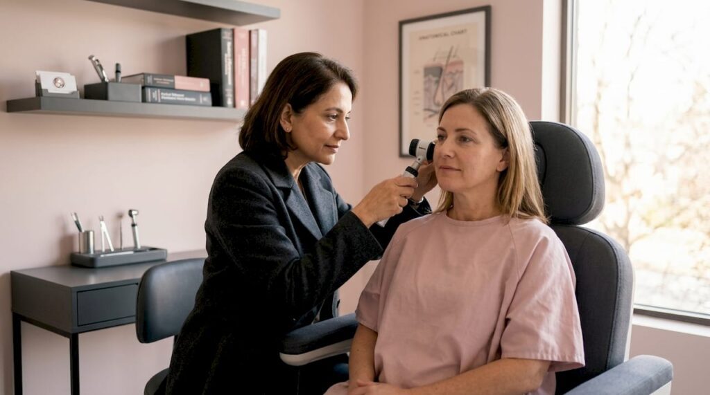
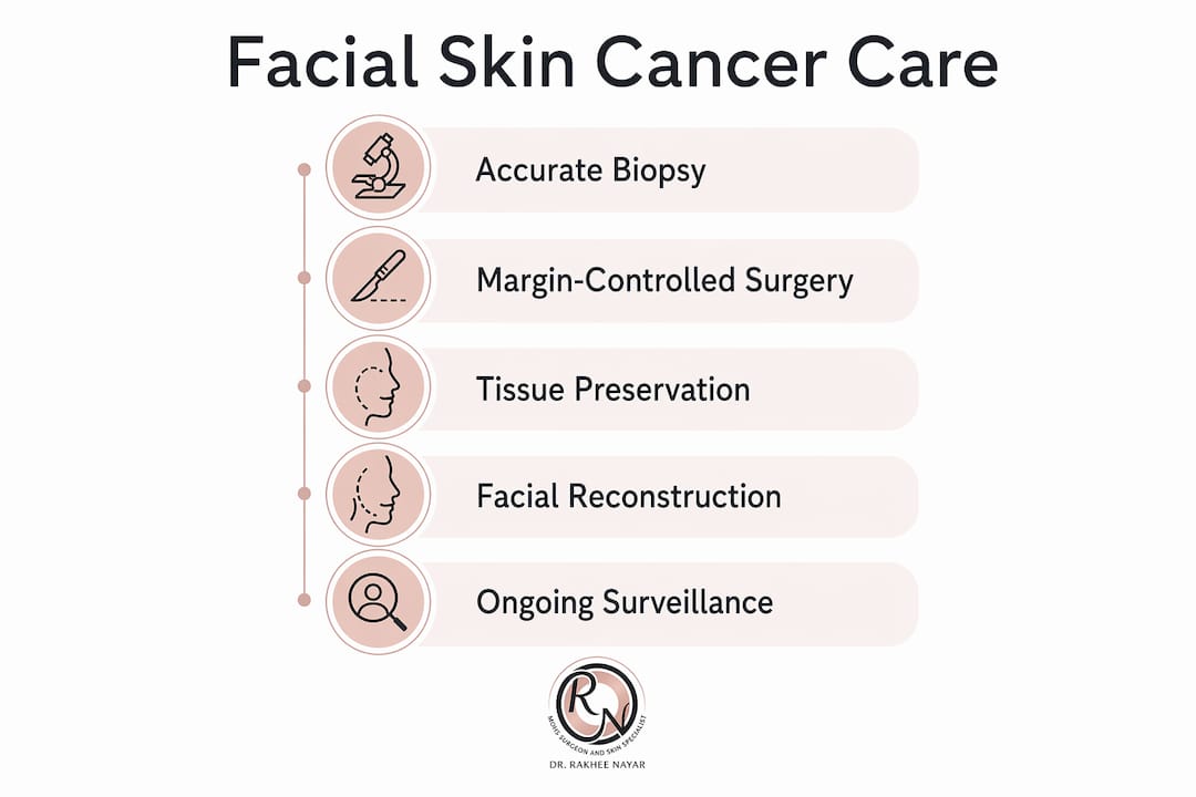
Facial skin cancer is defined as a malignant tumour arising on the face, where proximity to the eyes, nose, lips, and ears makes treatment far more demanding than anywhere else on the body. The reason why facial skin cancers need expert care comes down to three inseparable demands: complete tumour removal, preservation of healthy tissue, and reconstruction that restores both function and appearance. Standard excision techniques, used competently for cancers on the trunk or limbs, are simply not precise enough for the face. Specialist surgeons trained in margin-controlled surgery and facial reconstruction deliver measurably better outcomes, and the evidence behind that claim is clear.
Why facial skin cancers differ from other skin cancers
The face is not just cosmetically significant. It houses structures whose loss or damage carries serious functional consequences. The eyelids protect the cornea. The nose shapes airflow. The lips seal the mouth. Facial skin cancer involving these structures requires margin-controlled surgery to preserve function while achieving clear excision margins.
Standard surgical excision uses fixed margins, typically 4 to 6 mm for low-risk lesions and 6 to 10 mm for high-risk ones. On the trunk, removing that much tissue causes little disruption. On the face, the same approach can sacrifice tissue from an eyelid or the nasal ala that cannot be replaced without complex reconstruction. The risk cuts both ways: too little margin leaves residual tumour, too much causes unnecessary disfigurement.
High-risk histological subtypes compound this problem. Morphoeic basal cell carcinoma and poorly differentiated squamous cell carcinoma extend microscopically beyond their visible borders. A surgeon relying on clinical appearance alone will underestimate the true tumour extent in a significant proportion of cases.
Key reasons the face demands a different approach:
- Tumours near the eyelids, nose, and lips sit millimetres from structures that cannot be sacrificed
- High-risk subtypes spread subclinically, making visual margin assessment unreliable
- Cosmetic outcomes directly affect quality of life and psychological wellbeing
- Recurrent tumours on the face are harder to treat and carry higher morbidity
Pro Tip: If you are referred for skin cancer surgery on the face, ask specifically whether the surgeon has training in both Mohs micrographic surgery and facial reconstruction. These are distinct skill sets, and having both in one clinician changes the treatment plan from the outset.
How does biopsy technique affect facial skin cancer outcomes?
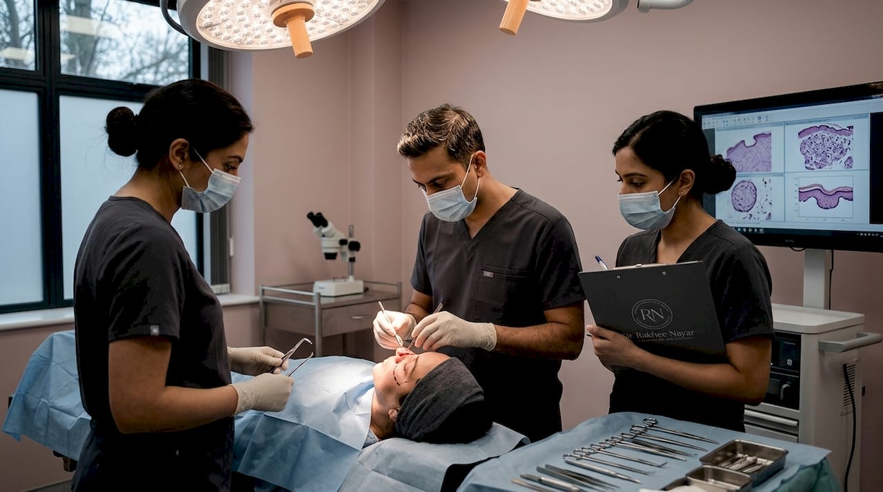
The quality of a biopsy determines the accuracy of everything that follows. An imprecise biopsy on the face does not just delay diagnosis. It can lead to incorrect staging, the wrong surgical plan, and ultimately a worse outcome.
Excisional biopsy with narrow margins of 1 to 3 mm is the preferred technique for suspected melanoma on the face. This approach removes the lesion intact, allowing the pathologist to assess the full Breslow thickness and determine the correct surgical margins for definitive treatment. Partial or incisional biopsies, which sample only a portion of the lesion, risk missing the deepest or most aggressive component entirely.
For basal cell carcinoma and squamous cell carcinoma, a punch or shave biopsy is often sufficient to confirm the diagnosis. However, the biopsy site location and technique still matter. A poorly placed biopsy can disrupt tissue planes and complicate subsequent Mohs surgery or reconstruction. Advanced imaging techniques such as reflectance confocal microscopy and optical coherence tomography are used in specialist settings to augment diagnosis and reduce unnecessary procedures, though biopsy remains the definitive step.
The practical implications for patients are straightforward:
- Insist on a biopsy before any surgical treatment begins
- Ask whether the biopsy technique is appropriate for the suspected cancer type
- Confirm that the biopsy report includes subtype, differentiation grade, and depth
- Understand that an incomplete or poorly planned biopsy may require a repeat procedure
What is Mohs surgery and why is it the gold standard?
Mohs micrographic surgery is a tissue-sparing, margin-controlled technique in which a surgeon removes the tumour in sequential horizontal layers, examining each layer under a microscope before proceeding. This continues until no cancer cells remain at any margin. The result is complete tumour clearance with the smallest possible defect.
The critical advantage lies in margin assessment. Standard vertical sectioning examines less than 1% of the surgical margin. Mohs surgery evaluates the complete peripheral and deep margin of every layer removed. This distinction is not academic. It means that microscopic tumour extensions, which would be missed by conventional histology, are identified and removed in the same procedure.
How a Mohs procedure works, step by step:
- The visible tumour is removed with a thin margin of surrounding tissue
- The tissue is mapped, colour-coded, and processed as frozen sections
- The Mohs surgeon examines 100% of the cut margins under the microscope
- If cancer cells remain at any margin, only that precise area is re-excised
- The cycle repeats until all margins are clear
- Reconstruction begins immediately once clearance is confirmed
| Feature | Standard excision | Mohs surgery |
|---|---|---|
| Margin assessment | Less than 1% of margin examined | 100% of peripheral and deep margin |
| Tissue preservation | Fixed margins removed regardless of tumour extent | Only tumour-positive areas re-excised |
| Same-day reconstruction | Rarely coordinated | Standard in specialist practice |
| Recurrence risk | Higher for high-risk facial tumours | Lower due to complete margin control |
Margin-controlled procedures remove cancer while sparing healthy tissue, directly reducing the extent of facial disfigurement and the complexity of subsequent reconstruction. For high-risk tumours on the face, the 2026 AAFP review identifies Mohs surgery as the treatment of choice, a position consistent with British Association of Dermatologists guidance.
Pro Tip: Mohs surgery is not appropriate for every facial skin cancer. It is most beneficial for high-risk subtypes, tumours in cosmetically sensitive locations, recurrent cancers, and lesions with poorly defined borders. A specialist assessment will determine whether it is the right choice for your situation.
What role does facial reconstruction play after skin cancer removal?
Removing a facial tumour creates a defect that must be repaired with the same level of precision as the excision itself. The reconstruction is not a secondary concern. It is an integral part of the treatment plan, and its quality directly determines the functional and cosmetic result.
Combining Mohs surgery and plastic reconstruction by a single dual-trained surgeon reduces delays and improves continuity of care. When excision and reconstruction are handled by separate teams, patients often wait days or weeks between procedures. That gap creates anxiety, leaves open wounds requiring temporary dressings, and means the reconstructive surgeon plans the repair without having been present during the excision.
The challenges of facial reconstruction are specific to location:
- Eyelid defects require techniques that maintain the blink reflex and protect the cornea
- Nasal reconstruction must restore the three-dimensional contour and preserve the airway
- Lip repairs need to maintain oral competence and natural movement
- Scalp and ear defects often require flap or graft techniques to cover exposed cartilage or bone
“Patients who have their Mohs surgery and reconstruction performed by the same surgeon on the same day consistently report lower anxiety, fewer appointments, and greater satisfaction with their cosmetic outcome. The continuity is not a luxury. It is a clinical advantage.”
Miss Rakhee Nayar offers same-day removal and reconstruction, closing the gap that exists when patients move between separate specialists. Her dual training in both Mohs surgery and plastic surgery means the reconstruction is planned with full knowledge of the tumour’s extent and the tissue available after clearance.
How to choose the right specialist for facial skin cancer
High-risk skin cancers on the face require referral to experienced clinicians who can optimise staging, excision, and reconstruction. Knowing what to look for in a specialist protects you from undertreatment and from unnecessary procedures.
The questions worth asking before committing to a surgeon:
- Are you trained in Mohs micrographic surgery and facial plastic reconstruction?
- What biopsy technique will you use, and will you review the pathology yourself?
- Do you perform same-day reconstruction, or will I be referred to a separate team?
- What is your policy on follow-up and ongoing skin surveillance?
Ongoing surveillance matters as much as the initial treatment. Patients who have had one facial skin cancer carry a significantly elevated risk of developing further lesions. A specialist who provides structured follow-up, rather than discharging you after surgery, offers a material long-term benefit.
Pro Tip: Ask to see before-and-after photographs of reconstructive outcomes for defects similar in location and size to yours. A surgeon confident in their results will share these readily.
Key takeaways
Expert care for facial skin cancer requires margin-controlled surgery, accurate biopsy technique, and integrated reconstruction to achieve the best cure rates and cosmetic outcomes.
| Point | Details |
|---|---|
| Margin control is non-negotiable | Mohs surgery assesses 100% of surgical margins, unlike standard excision which examines less than 1%. |
| Biopsy technique shapes the treatment plan | Excisional biopsy with narrow margins is preferred for suspected melanoma to avoid incomplete staging. |
| Facial anatomy demands specialist skill | Proximity to eyelids, nose, and lips means tissue loss carries functional as well as cosmetic consequences. |
| Dual-trained surgeons improve outcomes | Same-day Mohs surgery and reconstruction reduces delays, anxiety, and the risk of suboptimal repair. |
| Early specialist referral is critical | High-risk facial tumours require multidisciplinary pathways and structured follow-up to minimise recurrence. |
Expert Mohs surgery and facial reconstruction in the UK
If you have been diagnosed with a facial skin cancer, or you are concerned about a lesion that needs assessment, the most important step is a consultation with a specialist who can evaluate both the surgical and reconstructive needs together.

Rakhee Nayar – Mohs Surgeon and Skin Specialist offers Mohs micrographic surgery and same-day facial reconstruction from her clinic in North West England, with private consultations and e-consultations available for UK and international patients. Her dual training in Mohs surgery and plastic surgery means your treatment plan covers excision and repair as a single coordinated pathway. For a full overview of surgical options, the skin cancer treatment pages provide detailed guidance on what to expect at every stage.
FAQ
Why do facial skin cancers need specialist treatment?
Facial skin cancers sit adjacent to critical structures including the eyelids, nose, and lips, where standard excision margins are insufficient and tissue loss carries functional consequences. Specialist techniques such as Mohs surgery provide complete margin assessment while preserving the maximum healthy tissue.
What is the difference between Mohs surgery and standard excision?
Standard excision examines less than 1% of the surgical margin using vertical sectioning, whereas Mohs surgery maps and examines 100% of the peripheral and deep margin in real time. This makes Mohs significantly more accurate for high-risk or cosmetically sensitive facial tumours.
How does biopsy technique affect my treatment plan?
An accurate biopsy determines the cancer subtype, depth, and differentiation grade, all of which drive the surgical approach. Partial or incisional biopsies risk missing the most aggressive component of a tumour, leading to incorrect staging and an inadequate treatment plan.
What does facial reconstruction involve after skin cancer removal?
Facial reconstruction repairs the defect left after tumour excision using local flaps, skin grafts, or more complex techniques depending on the location and size of the wound. The goal is to restore both appearance and function, including movement, sensation, and structural integrity.
When should I seek a specialist opinion about a facial lesion?
Seek a specialist opinion promptly if a lesion on your face is growing, bleeding, failing to heal, or has an irregular border or colour. Early referral to a facial skin cancer specialist gives you the widest range of treatment options and the best chance of a straightforward outcome.
Recommended
- Types of skin cancer and advanced facial treatments UK
- High-risk skin cancer: Expert treatment guide for UK patients
- How to Spot Skin Cancer Symptoms on the Face – Expert Mohs Surgery | UK
- Expert plastic surgery in skin cancer care: best results
Filed under Choosing Your Care


