TL;DR:
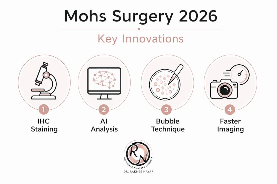
- Mohs surgery in 2026 now uses advanced techniques like AI-assisted margin assessment and IHC staining, reducing healthy tissue removal and improving accuracy for facial skin cancers. Innovations such as the bubble technique enhance tissue processing, while emerging imaging tools like CLSM offer faster, superficial margin evaluation, complementing traditional methods. Patients should inquire about these technologies and early reconstruction planning to achieve better outcomes and cosmetic results.
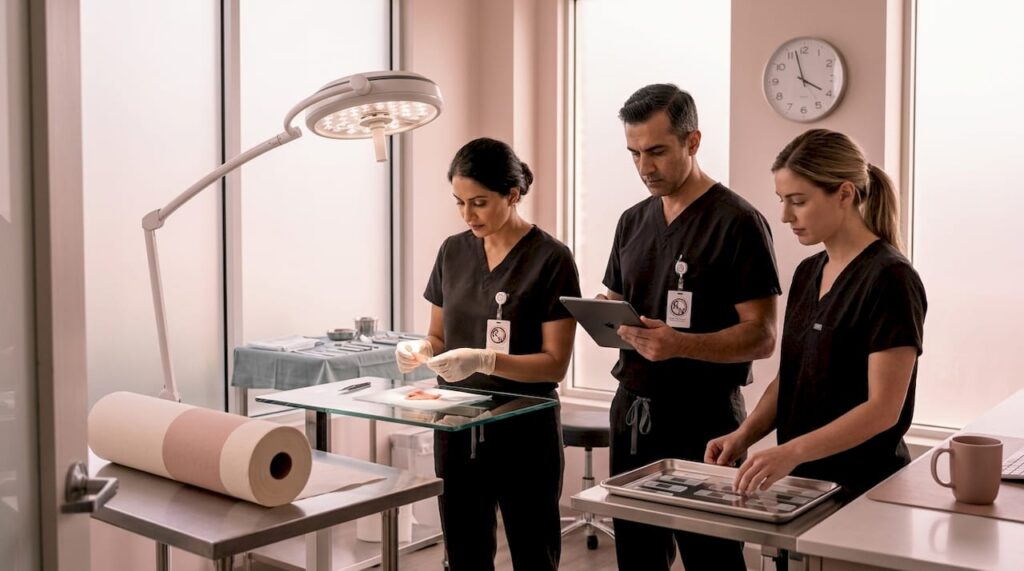
The latest Mohs surgery techniques in 2026 are doing something that would have seemed improbable a decade ago: they are making an already precise procedure even more accurate, while simultaneously reducing the amount of healthy tissue removed. For patients facing skin cancer on the face, that combination matters enormously. This article explains the specific advances reshaping Mohs surgery right now, from AI-assisted margin reading to a novel method called the bubble technique, so you can walk into any consultation knowing exactly what questions to ask and what to expect.
Table of Contents
- How Mohs surgery has advanced to treat melanoma and high-risk facial cancers
- Innovations in specimen processing: the bubble technique and conservative thickness layers
- Emerging diagnostic tools complementing Mohs surgery for faster margin evaluation
- What patients should know: questions to ask and what to expect from 2026 Mohs surgery techniques
- Why the latest Mohs surgery techniques signal a new era in skin cancer treatment
- Explore expert Mohs surgery options in the UK today
- Frequently asked questions
Key Takeaways
| Point | Details |
|---|---|
| Immunohistochemistry use | IHC staining in Mohs surgery highlights melanoma cells to ensure complete tumour removal during the procedure. |
| AI-supported precision | Artificial intelligence aids real-time margin analysis, improving accuracy and speed in Mohs surgery. |
| Bubble technique benefits | Specialised tissue processing improves slide quality and supports better cosmetic outcomes for delicate facial areas. |
| Complementary diagnostic tools | New imaging methods like CLSM complement Mohs surgery but don’t replace traditional margin evaluation yet. |
| Patient empowerment | Knowing the latest techniques helps patients ask informed questions and understand their treatment journey better. |
How Mohs surgery has advanced to treat melanoma and high-risk facial cancers
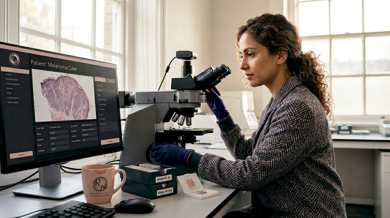
Building on the core principles of Mohs micrographic surgery, the most clinically significant shift in recent years involves how surgeons handle melanoma and high-risk facial lesions. Standard frozen-section analysis works well for basal cell carcinoma and squamous cell carcinoma, but melanoma cells are notoriously difficult to spot on routine slides. That is where immunohistochemistry (IHC) staining has changed the game.
IHC staining applies targeted chemical markers to tissue slides that bind specifically to melanoma cells, making them visible under a microscope even when standard processing would miss them. When IHC staining highlights cancer cells, the surgeon can detect residual tumour during the procedure itself, rather than discovering a positive margin days later. For facial melanoma, that real-time information is the difference between a single-stage excision and returning for further surgery.
Alongside IHC, artificial intelligence is beginning to support margin assessment in ways that matter to patients. AI-assisted margin assessment is a step toward real-time precision, improving both the speed and consistency of how slides are interpreted. The surgeon retains full decision-making authority, but AI reduces the variability that comes with human fatigue or the inherent subjectivity of reading complex slides.
The practical benefits for patients considering choosing Mohs surgery for facial skin cancer include:
- Fewer stages needed to achieve clear margins, particularly for melanoma
- Reduced removal of healthy tissue around the tumour site
- Lower likelihood of needing a second operation for residual cancer
- More accurate margin mapping for complex facial anatomy
“When dealing with melanoma on the face, IHC staining combined with en face margin assessment gives us information during the procedure that we simply cannot get from standard frozen sections alone.”
These mohs surgery advancements in 2026 are particularly relevant if you are managing a lesion near the eye, nose, ear, or lip, where tissue conservation directly shapes your long-term appearance and function. If you have been diagnosed with a high-risk or recurrent skin cancer, our high-risk skin cancer treatment guide explains what to expect from a specialist assessment.
Innovations in specimen processing: the bubble technique and conservative thickness layers
Beyond the diagnostic tools, advances in how tissue is physically prepared during Mohs surgery are proving equally important. The principle is straightforward: thinner tissue layers preserve more of the surrounding healthy anatomy. But processing those thin layers has traditionally created technical headaches that could compromise the accuracy of the entire procedure.
When a tissue layer is extremely thin, it tends to fold, curl, and separate during slide preparation. Those deformations create gaps and artefacts that can obscure the true surgical margin, potentially masking residual tumour cells. In practice, this has sometimes pushed Mohs histotechnicians towards slightly thicker layers to guarantee slide quality, which works against the goal of tissue conservation.
The bubble technique resolves this tension directly. Here is how it works in sequence:
- The freshly excised tissue layer is placed on the slide in the standard orientation.
- A small frozen bead of embedding medium is placed on top of the tissue before full freezing occurs.
- As the medium freezes, it creates gentle, even pressure that flattens the epidermis against the slide surface.
- The result is a flatter, more uniform tissue section that allows comprehensive evaluation of conservative thickness layers, improving both slide quality and margin assessment accuracy.
This may sound like a minor laboratory refinement, but its clinical significance is real. Better slide quality means the surgeon can read margins with greater confidence. Greater confidence means fewer precautionary additional stages. Fewer stages means less tissue removed and a simpler Mohs surgery recovery workflow overall.
Pro Tip: When meeting your surgeon, ask not only about the surgical technique but also about how tissue processing is handled at the facility. The quality of slide preparation directly affects how accurately your margins can be assessed.
The bubble technique is one reason why new mohs techniques in 2026 are worth knowing about even if you have already read general guides on the procedure. It represents the kind of incremental but meaningful refinement that reinforces why Mohs is the gold standard for skin cancer removal on the face.
Emerging diagnostic tools complementing Mohs surgery for faster margin evaluation
Alongside improvements in specimen processing, newer imaging technologies are beginning to support the Mohs workflow with real-time tumour margin information that does not require traditional slide preparation at all.
Ex vivo confocal laser scanning microscopy (CLSM) is the most clinically advanced of these approaches. The technique works by scanning freshly excised tissue with a focused laser, producing images that closely resemble conventional histology without the time and processing steps involved in standard frozen sections. The key advantage is speed: CLSM can generate margin images within minutes of excision, potentially compressing the waiting time between surgical stages.
However, the technology has genuine limitations. Ex vivo CLSM provides real-time quasi-histological visualisation of tumour architecture and margins, but its penetration depth is approximately 250 micrometres. That restriction means it cannot assess the full depth of thicker lesions, and it works best for superficial tumour types. It complements frozen-section Mohs analysis rather than replacing it, at least for now.
The following table summarises how CLSM compares with conventional Mohs frozen-section analysis across the factors that matter most to patients:
| Feature | Conventional frozen sections | Ex vivo CLSM |
|---|---|---|
| Imaging speed | 45 to 90 minutes per stage | 5 to 20 minutes per stage |
| Tissue processing required | Yes | No |
| Penetration depth | Full thickness | Approximately 250 µm |
| Best suited to | All skin cancers | Superficial lesions |
| Current role in Mohs | Standard practice | Complementary tool |
For patients with a superficial basal cell carcinoma or thin squamous cell carcinoma, combining CLSM with standard processing could shorten the overall procedure duration and reduce time spent waiting between stages. Better skin cancer mapping techniques at every stage of the workflow translate into a more comfortable and less disruptive day for you.
The future of Mohs surgery will likely involve CLSM and similar tools becoming standard adjuncts in specialist centres, particularly as the technology becomes more accessible and evidence accumulates.
What patients should know: questions to ask and what to expect from 2026 Mohs surgery techniques
With the technical and diagnostic advances explained, the most practical question is: how do these changes affect your specific care journey? Understanding that is the difference between passively receiving treatment and actively participating in decisions about your own surgery.
Here are the key things to confirm before and during your consultation:
- Ask whether IHC staining is used. Patients should ask if their surgeon uses IHC staining and en face margin assessment for melanoma, because not every facility offers it and it significantly affects how thoroughly margins can be assessed in real time.
- Understand that thin-layer processing may mean fewer stages. If your surgeon uses the bubble technique or similar methods, conservative tissue removal becomes more reliably achievable, which tends to simplify post-surgical reconstruction.
- Expect longer waits between stages for melanoma. IHC staining takes additional time compared with standard frozen sections. That is not a sign something has gone wrong. It is a sign the most thorough approach is being used.
- Discuss reconstruction early, not after. Knowing your facial reconstruction options before surgery, rather than after, allows your surgical team to plan excision and closure together, which generally leads to better cosmetic results.
Pro Tip: If your lesion is on the face and you have been offered a choice between standard excision and Mohs surgery, reviewing the evidence on Mohs versus standard excision with your surgeon will clarify why Mohs consistently achieves higher cure rates for high-risk or recurrent facial cancers.
Recovery time varies considerably depending on the size of the defect and whether a flap, graft, or direct closure is used. Your surgeon should explain this before you leave the consultation, not on the day of the procedure.
Why the latest Mohs surgery techniques signal a new era in skin cancer treatment
Here is something worth saying plainly: the 2026 mohs surgery updates described above are not just incremental improvements to a static procedure. They represent a shift in what the procedure actually is.
For most of its history, Mohs surgery was defined by what it removed and how thoroughly. The measure of success was a clear margin. That remains true. But the mohs surgery innovations in 2026 are adding a second dimension: how little healthy tissue is sacrificed in the process of achieving that clear margin. The bubble technique, IHC staining, and AI-assisted reading are not about doing the same thing faster. They are about doing something qualitatively different, where the precision of removal is matched by the intelligence of the diagnostic process.
The integration of AI in Mohs workflows represents a shift towards real-time precision and consistency in margin assessment that was simply not possible with purely manual interpretation. That matters especially for facial surgery, where a single additional millimetre of unnecessary excision near the eyelid or nasal ala can mean a significantly more complex reconstruction.
From a clinical perspective, the patients who benefit most from these advances are those with melanoma on cosmetically sensitive areas, recurrent tumours where previous surgery has already disrupted local tissue architecture, and lesions at the boundaries of vital facial structures. These are exactly the cases where Mohs as the gold standard has always been most justified, and where the new techniques deliver their clearest advantages.
The direction of travel in this field is consistent: more information, gathered sooner, interpreted more reliably, acting in service of the best possible outcome for both tumour clearance and appearance. The mohs surgery best practices of 2026 are the foundation for what will become routine care within the next five to ten years.
Explore expert Mohs surgery options in the UK today
If you are facing a diagnosis of skin cancer on the face or another cosmetically sensitive area, accessing care that incorporates these latest advances makes a genuine difference to your outcome.
Miss Rakhee Nayar offers specialist Mohs surgery with dual training in both Mohs micrographic surgery and plastic surgery, a combination that means your tumour removal and facial reconstruction are planned together from the start. You can explore thorough skin cancer detection services to understand your diagnosis fully before treatment begins. When you are ready to discuss surgical options, the Mohs surgery services page outlines exactly what the procedure involves and what to expect. For patients who need post-surgical repair, personalised facial reconstruction ensures both function and appearance are restored with the same level of expertise.
Frequently asked questions
What is immunohistochemistry (IHC) and why is it important in Mohs surgery for melanoma?
IHC is a staining technique applied to tissue slides during Mohs surgery that chemically highlights melanoma cells, making them visible under the microscope even when standard processing would miss them. Because IHC staining detects residual tumour during the procedure itself, the surgeon can achieve clear margins in real time rather than discovering a problem days later.
How does AI assist margin assessment in Mohs surgery?
AI analyses surgical margin slides quickly and consistently, supporting the surgeon’s interpretation with standardised pattern recognition that reduces the variability of manual reading. The technology means AI-assisted margin interpretation improves both the speed and accuracy of decisions about how much tissue to remove.
What is the bubble technique in Mohs surgery specimen processing?
The bubble technique places a frozen bead of embedding medium onto thin tissue sections during slide preparation, creating even pressure that flattens the tissue and prevents folding or separation. The outcome is that the bubble technique improves slide quality and the accuracy of margin assessment for conservative, thin-layer excisions.
Can newer imaging tools replace traditional Mohs surgery margin analysis?
Not currently. Ex vivo CLSM produces rapid, detailed margin images but its penetration depth of 250 µm limits its use to superficial lesions, meaning it works as a complement to frozen-section Mohs analysis rather than a replacement for established practice.