Melanoma spots are abnormal pigmented skin lesions that arise from melanocytes, the cells that give skin its colour. They are the most serious form of skin cancer, and recognising them early is the single most important factor in improving treatment outcomes. UK melanoma cases are projected to reach 26,500 annually by 2040, a figure that reflects decades of cumulative UV damage across the population. Miss Rakhee Nayar, GMC-registered Consultant Plastic Surgeon and Mohs specialist, directs the clinical guidance throughout this article to help you understand what to look for, where to look, and when to seek expert review.
1. What do melanoma spots look like?
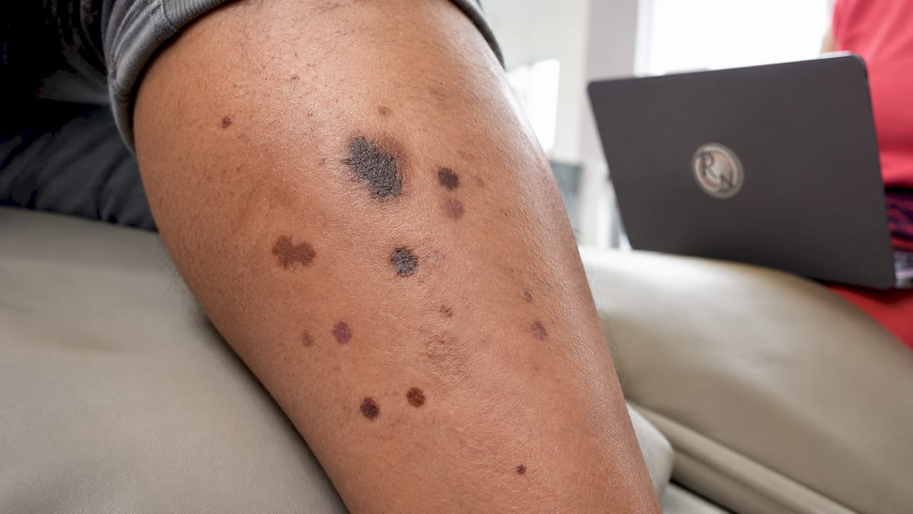
Melanoma spots, known clinically as malignant melanoma lesions, share several recognisable features that distinguish them from ordinary moles or freckles. The ABCDE rule is the most widely accepted clinical guide for assessing suspicious pigmented lesions.
- Asymmetry. One half of the spot does not match the other.
- Border. The edges are irregular, ragged, notched, or blurred rather than smooth and round.
- Colour. The lesion contains more than one shade, including brown, black, red, white, or blue within the same spot.
- Diameter. Most melanomas are larger than 6mm across, roughly the size of a pencil eraser, though some are smaller when first detected.
- Evolving. Any change in size, shape, colour, or texture over weeks or months is a warning sign.
Early-stage melanoma may not cause any symptoms at all. That absence of pain is one reason people delay seeking advice. Beyond appearance, some lesions produce tingling, itching, bleeding, or crusting before other visible changes become obvious.
Pro Tip: Photograph any suspicious spot monthly under the same lighting conditions. Side-by-side comparison over 8–12 weeks reveals subtle changes that are easy to miss in daily life.
2. Where on the body do melanoma spots most commonly appear?
Melanomas can appear anywhere on the skin, but the most frequently affected sites differ between men and women. Men develop melanomas most often on the torso, particularly the back. Women are more commonly affected on the legs.
Beyond these common sites, several areas are frequently overlooked during self-examination:
- The scalp, particularly along the hairline and parting
- The ears, including the outer rim and behind the ear
- The soles of the feet and between the toes
- Under and around the fingernails and toenails
- The lower back and buttocks
These hidden locations matter because melanomas in less visible areas tend to be diagnosed at a later stage. Later diagnosis means more advanced disease and more complex treatment.
Nearly 9 out of 10 melanoma cases in the UK are caused by excessive UV radiation from sun exposure and sunbeds. That said, melanomas can also develop on skin that receives little or no sun exposure, including the palms, soles, and mucous membranes. This means UV protection alone does not eliminate risk entirely, and whole-body vigilance is necessary.
3. How to tell melanoma spots apart from benign skin marks
Distinguishing a melanoma from a harmless skin mark is not always straightforward, even for experienced clinicians. The ABCDE system provides a practical starting framework, but several benign conditions can mimic the appearance of melanoma.
Common benign lookalikes include:
- Freckles (ephelides). Small, flat, uniformly brown spots that fade in winter and are common in fair-skinned people. They are symmetrical and stable.
- Seborrhoeic keratoses. Waxy, stuck-on-looking growths that are often brown or grey. They can be mistaken for melanoma because of their irregular surface, but they do not arise from melanocytes.
- Solar lentigines. Flat, evenly pigmented brown spots caused by cumulative sun exposure. They are common in adults over 40 and are generally stable in appearance.
The features that suggest a lesion needs urgent review are different from the standard ABCDE criteria. Nodular melanoma can present as a fast-growing firm bump or flat lesion that bleeds or crusts. It does not always follow the ABCDE pattern, which is why any new lump that changes rapidly warrants prompt medical attention regardless of colour or shape.
Pro Tip: If a lesion looks like a seborrhoeic keratosis but has appeared recently and is growing, treat it as suspicious. Seborrhoeic keratoses are stable lesions; new, growing ones deserve specialist review.
Satellite moles, which are small pigmented spots appearing near an existing lesion, are another sign that warrants immediate referral. They can indicate local spread of melanoma cells through the lymphatic system.
4. What are the signs of melanoma spots that go beyond appearance?
Visual assessment alone does not capture every warning sign. Early symptoms like itching or tingling in a spot can precede visible changes, which means self-awareness extends beyond what you can see.
Tactile and symptomatic changes to watch for include:
- Persistent itching in a mole or pigmented spot that has not previously been itchy
- A sensation of tingling or warmth within a lesion
- Bleeding that occurs without injury
- Crusting or scabbing that repeatedly returns after healing
- A feeling that the skin surface over a spot has changed in texture
These symptoms do not confirm melanoma on their own. They do, however, indicate that a lesion is biologically active in some way, and that warrants clinical assessment. Waiting to see whether a symptom resolves is not the appropriate response when a pigmented lesion is involved.
You can read more about early detection benefits and why acting on early symptoms significantly improves outcomes.
5. How to examine your skin for suspicious spots
A systematic approach to self-examination is the most reliable way to detect changes early. Using mirrors and assistance helps to check areas that are difficult to see unaided.
Follow these steps monthly:
- Examine your face. Use a well-lit mirror to check your nose, lips, ears, and scalp along the hairline.
- Check your scalp. Use a comb or hairdryer to part the hair in sections. Ask someone to help if possible.
- Inspect your hands. Look at your palms, the backs of your hands, between your fingers, and under your nails.
- Examine your arms and torso. Raise each arm and use a full-length mirror to check your underarms, chest, and abdomen.
- Check your back and buttocks. Use a hand mirror in front of a full-length mirror to see your back, shoulders, and the back of your neck.
- Inspect your legs and feet. Sit down to examine the backs of your legs, the soles of your feet, between your toes, and under your toenails.
When to seek medical advice is straightforward: any lesion that meets one or more ABCDE criteria, any new or changing spot, or any lesion causing symptoms should be assessed by a clinician without delay. A regular full body check-up with a healthcare professional adds an important layer of detection beyond self-examination.
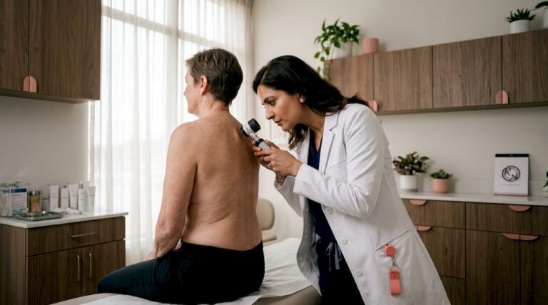
Specialists use dermoscopy, a technique that magnifies and illuminates the skin surface, to assess lesions in detail that the naked eye cannot resolve. When dermoscopy raises concern, a biopsy is taken and the tissue is examined histologically to confirm or exclude melanoma. This is the definitive diagnostic step.
6. What can you do to reduce your risk of developing melanoma spots?
Prevention reduces risk but does not eliminate it entirely. The most effective approach combines multiple strategies rather than relying on any single measure.
“Avoiding sunburn is the most critical prevention strategy against melanoma. Recommendations include seeking shade between 11 AM and 3 PM, wearing protective clothing, and applying broad-spectrum sunscreen with SPF 30 or higher and a 4 or 5 star UVA rating. Reapplication every two hours, and after swimming or sweating, is necessary for sustained protection.”
Sun safety practices are most effective when combined: shade, protective clothing, and sunscreen together provide substantially better protection than any one measure alone.
Key prevention habits include:
- Seek shade between 11 AM and 3 PM, when UV intensity is highest.
- Wear protective clothing, including long sleeves, wide-brimmed hats, and UV-protective sunglasses.
- Apply SPF 30+ broad-spectrum sunscreen to all exposed skin, including the ears, back of the neck, and tops of the feet.
- Reapply sunscreen every two hours and immediately after water exposure.
- Avoid sunbeds entirely. Artificial UV sources carry the same melanoma risk as natural sunlight and are not safer.
- Attend regular skin checks with a specialist, particularly if you have a personal or family history of melanoma, fair skin, or a large number of moles.
UV rays penetrate cloud cover, which means sun protection is necessary even on overcast days. Many patients are surprised to learn that significant UV damage accumulates during everyday activities such as driving or gardening, not only during deliberate sunbathing.
You can find further guidance on skin health practices that support both prevention and recovery after treatment.
Key takeaways
Early recognition of melanoma spots is the most effective way to improve treatment outcomes and reduce the risk of advanced disease.
| Point | Details |
|---|---|
| Use the ABCDE rule | Asymmetry, Border, Colour, Diameter, and Evolving changes are the primary indicators of a suspicious lesion. |
| Check hidden areas | The scalp, ears, feet, and under nails are frequently missed during self-examination but carry real risk. |
| Symptoms matter | Itching, tingling, or bleeding in a pigmented spot can precede visible changes and should prompt clinical review. |
| Combine prevention strategies | Shade, protective clothing, and SPF 30+ sunscreen together offer the best protection against UV damage. |
| Seek specialist assessment early | Any changing, symptomatic, or rapidly growing lesion warrants prompt review by a GMC-registered specialist. |
What I see in clinic that patients often miss
Patients frequently arrive at my clinic having watched a lesion change for six months or more before seeking advice. The most common reason they give is uncertainty: they were not sure whether what they were seeing was “bad enough” to warrant a consultation. That hesitation is understandable, but it is also the single biggest obstacle to early diagnosis.
The ABCDE rule is a useful guide, but it has real limitations. Nodular melanoma, in particular, can be fast-growing and aggressive without displaying the classic irregular border or multiple colours that patients expect. I have seen lesions dismissed as cysts or insect bites that turned out to be nodular melanoma. A firm, new lump that was not there three months ago deserves clinical attention, full stop.
Another pattern I notice is that patients check the areas they can see easily and neglect the rest. The back, scalp, and soles of the feet are consistently under-examined. If you cannot see an area clearly, ask a partner or family member to help, or arrange a professional skin check. The few minutes it takes are genuinely worthwhile.
The myth that melanoma only affects people who sunbathe is also worth addressing directly. Acral lentiginous melanoma, which develops on the palms, soles, and under nails, has no established link to UV exposure. It is less common but no less serious. Whole-body awareness is the only approach that covers all presentations.
Acting early is not overcaution. It is the clinically correct response to a changing or symptomatic skin lesion. If you are uncertain, a consultation is always the right next step.
— Miss Rakhee Nayar
Concerned about a suspicious spot? Expert assessment is available
If you have noticed a changing mole, a new pigmented lesion, or a spot that does not look quite right, a specialist consultation is the appropriate next step. Rakhee Nayar – Mohs Surgeon and Skin Specialist offers expert skin cancer assessment and treatment at a private clinic in North West England, with e-consultations available for patients across the UK and internationally.

Miss Nayar is dual-trained in plastic surgery and Mohs micrographic surgery, which means she assesses lesions with both oncological and reconstructive expertise from the outset. For patients whose lesions require surgical treatment, Mohs micrographic surgery offers precise removal with the highest cure rates available for skin cancer. You can also review skin cancer symptoms in detail before your appointment. To book a private consultation, visit mohssurgeon.co.uk.
This article is for information only and does not constitute medical advice. Consult a GMC-registered specialist for assessment of any suspicious skin lesion.
FAQ
What are the first signs of melanoma on the skin?
The first signs are often a change in an existing mole or the appearance of a new pigmented spot. Changes in colour, shape, or size, and symptoms such as itching or tingling, are early indicators that warrant clinical review.
How is melanoma different from a normal mole?
A normal mole is symmetrical, has a smooth border, is a single shade of brown, and remains stable over time. A melanoma lesion typically shows asymmetry, irregular borders, multiple colours, a diameter greater than 6mm, or visible change over weeks or months.
Can melanoma spots appear on areas not exposed to the sun?
Yes. Melanoma can develop on the palms, soles, under nails, and mucous membranes, areas with little or no UV exposure. This form, known as acral lentiginous melanoma, is less common but requires the same prompt assessment as sun-related melanoma.
How often should I check my skin for suspicious lesions?
A monthly self-examination is recommended, combined with an annual professional skin check, particularly for adults over 40, those with fair skin, or those with a personal or family history of skin cancer.
When should I see a specialist rather than my GP?
See a specialist promptly if a lesion is growing rapidly, bleeding without injury, or does not fit the typical appearance of a benign mole. A GMC-registered skin cancer specialist can perform dermoscopy and arrange biopsy if needed, providing a definitive assessment that a GP referral pathway may delay.