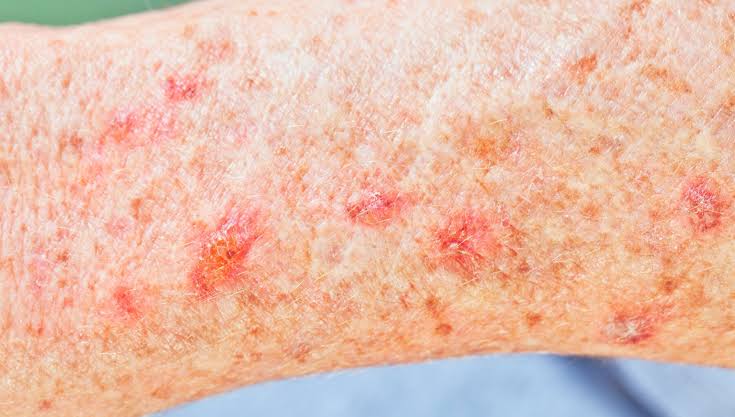
A white skin cancer spot is a lesion most commonly associated with basal cell carcinoma (BCC) or squamous cell carcinoma (SCC), presenting as a pearly, waxy, shiny, or scar-like white area on the skin. The clinical term for this presentation is a non-pigmented or hypopigmented skin malignancy. These spots are frequently mistaken for benign conditions such as milia, sebaceous hyperplasia, or simple scarring, which is precisely why they carry a higher risk of delayed diagnosis. Recognising a white skin cancer spot early is the single most important factor in achieving a straightforward, low-risk treatment outcome. This article explains what to look for, how specialists confirm the diagnosis, and which treatment options are used, including Mohs micrographic surgery.
If you have a spot like this. A white or pearly patch that is not healing needs a specialist opinion rather than watchful waiting. See the skin cancer treatment and surgery options, or arrange a consultation with Miss Nayar.
White spots on the skin: the short answers
What does a cancerous white spot look like?
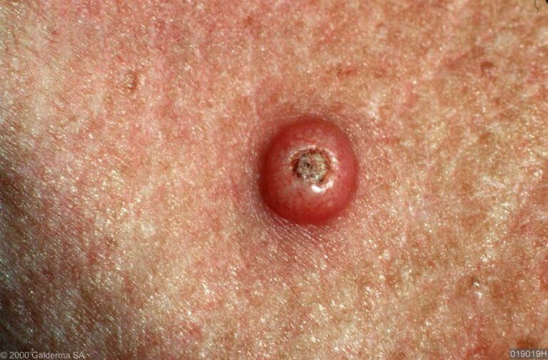
A cancerous white spot is usually pearly or waxy rather than flat and chalky, and it often has a raised, translucent edge. Fine blood vessels may run across the surface. It may bleed after a knock, scab over, then break down again in the same place.
How can you tell if a white spot is skin cancer?
By whether it changes. Harmless white spots such as milia stay the same for years. A cancer grows, spreads outwards, alters in texture, or repeatedly fails to heal. Any white lesion still present after four weeks, or changing at all, should be examined by a specialist.
What kind of cancer causes white spots on the skin?
Most often basal cell carcinoma, and less often squamous cell carcinoma. Both can look white, pearly or scar-like when they carry no pigment. Melanoma is usually dark, though a rare amelanotic form can appear pale or pink (NHS, symptoms of non-melanoma skin cancer).
When should you worry about white patches on the skin?
When a patch bleeds or crusts repeatedly, has a raised pearly rim, is growing, or sits on the face, scalp or ears. Widespread flat white patches with no raised edge are more likely to be a pigment condition such as vitiligo than a cancer.
What causes white skin cancer spots and who is at risk?
Cumulative ultraviolet (UV) radiation exposure is the primary cause of both basal cell carcinoma and squamous cell carcinoma, the two cancers most likely to produce white or pearly lesions. UV damage accumulates over decades, which is why these cancers appear most frequently in adults aged 40–75. The face, scalp, ears, neck, and backs of the hands are the sites most commonly affected, as these areas receive the greatest lifetime sun exposure.
Several factors increase individual risk significantly:
- Fair skin and light colouring. People with pale skin, light eyes, and a tendency to burn rather than tan carry the highest baseline risk.
- Age over 50. The majority of BCCs and SCCs are diagnosed in this age group, though younger adults with high UV exposure are not exempt.
- Immunosuppression. Organ transplant recipients and patients on long-term immunosuppressive therapy face a substantially elevated risk of SCC in particular.
- Previous skin cancer history. A prior BCC or SCC significantly raises the likelihood of a further lesion developing within five years.
- Genetics. Conditions such as Gorlin syndrome (basal cell naevus syndrome) predispose individuals to multiple BCCs from an early age.
- Chronic wounds or scars. SCC can arise within areas of long-standing scarring or chronic skin inflammation.
Benign conditions including milia (small white keratin cysts) and sebaceous hyperplasia (enlarged sebaceous glands) can appear visually similar to early BCCs. The critical difference is that malignant lesions change over time. Benign spots tend to remain stable for years, whereas a cancerous white spot typically evolves in size, texture, or surface character. UV damage remains the common thread, and anyone with a history of significant sun exposure should treat any new or changing white lesion as suspicious until proven otherwise.
How to identify white skin cancer spots: recognising symptoms and signs
Basal cell carcinoma is the most common form of skin cancer and typically appears as a small, shiny, firm bump with a pearly white or translucent border, sometimes with a central depression or ulceration. The surface may show fine blood vessels threading across it, and the lesion can bleed easily after minor trauma. SCC, by contrast, often begins as a rough, scaly patch that gradually develops into a raised spot or open sore that bleeds intermittently.
The following features should prompt urgent professional evaluation:
- A white, pearly, or shiny spot that has appeared or changed within the past few weeks to months
- A sore that does not heal within four weeks
- A flat, pale, scar-like area with no clear history of injury
- A lesion with an irregular or rolled border
- Visible fine blood vessels (telangiectasias) crossing the surface of the spot
- Any spot that bleeds, crusts, or weeps repeatedly
The ABCDE rule, familiar from melanoma awareness, applies in a modified form here. The ‘E’ for Evolving is the primary warning sign for non-pigmented lesions. A white spot that is growing, changing shape, or altering in texture demands assessment regardless of its colour. Colour alone is not a reliable guide.
Amelanotic melanoma is a rarer but serious consideration. It lacks melanin pigment and can appear white, pink, or skin-toned, making it visually indistinguishable from a BCC or a benign lesion without specialist examination. This is the most dangerous mimic in this category.
Pro Tip: Photograph any suspicious white spot in good natural light once a week. A side-by-side comparison over four to six weeks will reveal subtle changes in size or surface that are easy to miss in daily observation.
Common benign mimics include milia, dermatofibromas, and hypertrophic scars. None of these will evolve rapidly or bleed spontaneously. If a spot does either, treat it as suspicious.
How are white skin cancer spots diagnosed?
Accurate diagnosis of a white or pearly skin lesion follows a structured clinical pathway. Skipping steps in this process is the most common reason for delayed or missed diagnoses.
- Clinical examination. A specialist examines the lesion under direct lighting, assessing its border, surface texture, colour variation, and relationship to surrounding skin. The history of the lesion, including how long it has been present and whether it has changed, is equally important.
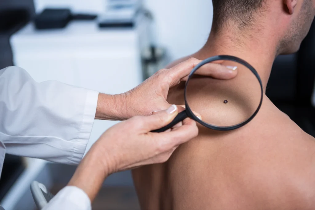
- Dermoscopy. A dermatoscope magnifies the lesion and reveals structures invisible to the naked eye. Dermoscopic evaluation detects telangiectasias, leaf-like structures, and spoke-wheel patterns that are characteristic of BCC. This step significantly increases diagnostic accuracy before any tissue is removed.
- Skin biopsy. Biopsy is the gold standard for definitive diagnosis. Biopsy distinguishes benign from malignant white lesions with certainty. A small sample of tissue is taken under local anaesthetic and sent for histopathological analysis. The result confirms the cancer type, subtype, and depth of invasion, all of which guide treatment planning.
- Histopathology review. The pathologist’s report identifies the specific BCC subtype (nodular, superficial, morphoeic) or SCC grade. Morphoeic BCC, which presents as a flat, scar-like white plaque, is particularly aggressive and requires wider surgical margins.
- Urgent referral criteria. Any lesion that is rapidly enlarging, located near the eye, nose, or ear, or suspected to be amelanotic melanoma warrants urgent referral under the NHS two-week-wait pathway or immediate private consultation.
Pro Tip: Ask your GP or specialist specifically whether dermoscopy was used during your assessment. A visual-only examination of a white lesion is insufficient for confident diagnosis.
Amelanotic melanoma challenges the classical ABCDE rules because it lacks the dark pigmentation that most people associate with melanoma. Persistence and evolution are more reliable indicators than colour in this context. Any white or pink spot that has been present for more than four weeks without explanation deserves a biopsy.
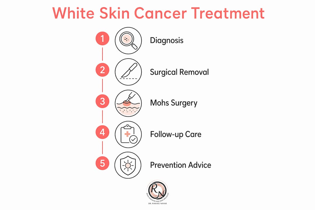
What are the treatment options for white skin cancer spots?
Treatment selection depends on the cancer type, subtype, anatomical location, lesion size, and the patient’s overall health. The table below summarises the main options.
| Treatment | Best Suited For | Key Advantage | Limitation |
|---|---|---|---|
| Mohs micrographic surgery | BCC and SCC on face, ears, nose, eyelids | Margins checked during surgery, tissue-sparing | Requires specialist centre |
| Standard surgical excision | Low-risk BCC or SCC on trunk or limbs | Widely available, straightforward | Fixed margins, higher recurrence risk for complex subtypes |
| Curettage and cautery | Superficial BCC only | Quick, minimal equipment | Not suitable for aggressive subtypes or high-risk sites |
| Radiotherapy | Patients unfit for surgery | Non-invasive | Lower cure rate than surgery, multiple sessions required |
| Topical treatments (e.g. imiquimod, 5-FU) | Superficial BCC only | Non-surgical | Not appropriate for nodular or morphoeic BCC |
Mohs micrographic surgery is the treatment of choice for BCCs and SCCs on the face, scalp, ears, and other cosmetically or functionally sensitive areas. The procedure removes the tumour in staged layers, with each layer examined under a microscope before the next is taken. This means the surgeon removes only cancerous tissue, leaving the maximum amount of healthy skin intact.
Surgical excision for skin cancer on the trunk or limbs remains the standard approach for lower-risk lesions. Standard excision uses fixed margins based on tumour type and size, without real-time margin assessment. For morphoeic BCC or recurrent SCC, this approach carries a meaningfully higher recurrence risk compared with Mohs surgery.
Mohs surgery is the gold standard for tumours in locations where tissue conservation matters, such as the nose, eyelid, and lip. For recurrent BCC, where standard excision has already failed, Mohs surgery remains the most reliable option.
Post-treatment follow-up is not optional. Patients treated for BCC or SCC should have annual skin checks for at least five years. The risk of a second primary skin cancer is significant in this population, and early detection of any new lesion keeps treatment options straightforward.
Key takeaways
A white or pearly skin lesion that evolves, bleeds, or fails to heal within four weeks requires specialist assessment and biopsy, not watchful waiting.
| Point | Details |
|---|---|
| White spots can be malignant | Basal cell carcinoma and squamous cell carcinoma both present as white, pearly, or shiny lesions. |
| Benign mimics are common | Milia and sebaceous hyperplasia look similar; only biopsy confirms the diagnosis with certainty. |
| Evolution is the key warning sign | Any white lesion that changes in size, texture, or surface within weeks warrants urgent evaluation. |
| Mohs surgery checks every margin | For facial and high-risk lesions, Mohs micrographic surgery checks every margin under the microscope and removes as little tissue as possible. |
| Annual follow-up is necessary | Patients with a history of BCC or SCC face elevated risk of further lesions and need regular skin checks. |
Concerned about a white spot? get a specialist opinion
Rakhee Nayar – Mohs Surgeon and Skin Specialist offers consultant-led assessment, dermoscopy, and biopsy for suspicious white skin lesions at Circle Cheshire in North West England. Miss Nayar holds dual training in plastic surgery and Mohs micrographic surgery, providing both precise cancer removal and considered reconstruction in a single specialist pathway. Private consultations and e-consultations are available for UK and international patients. If you have noticed a white, pearly, or changing spot on your skin, the right step is a formal assessment, not continued observation. Visit the skin cancer symptoms guide for detailed information on what to look for, or book a consultation directly through mohssurgeon.co.uk.
This article is for educational purposes only and does not constitute medical advice. Consult a GMC-registered specialist for assessment of any suspicious skin lesion.
FAQ
What does a white skin cancer spot look like?
A white skin cancer spot typically appears as a pearly, shiny, or waxy bump with a rolled or translucent border, sometimes with fine blood vessels visible on its surface. It may also present as a flat, pale, scar-like area that has appeared without a history of injury.
Can skin cancer be white rather than dark?
Yes. Basal cell carcinoma and squamous cell carcinoma both present as white or skin-coloured lesions. Amelanotic melanoma is a rarer but serious form that also lacks dark pigmentation, appearing white or pink.
How is a white skin lesion diagnosed as cancer?
Diagnosis requires a skin biopsy examined by a pathologist. Dermoscopy by a trained specialist can identify suspicious features before biopsy, but tissue analysis is the only definitive method for confirming malignancy.
Is mohs surgery suitable for white skin cancer spots on the face?
Mohs micrographic surgery is the preferred treatment for BCC and SCC on the face, nose, eyelids, and ears. Each margin is checked under the microscope during the procedure, before the wound is closed, so only the tissue needed to clear the cancer is removed. Read more about facial skin cancer removal to understand why Mohs is the preferred approach in these locations.
When should i see a specialist about a white spot on my skin?
Seek specialist assessment if a white spot has been present for more than four weeks, has changed in size or texture, bleeds or crusts repeatedly, or appears on the face, scalp, or ears. Do not wait for the lesion to become painful or visibly larger before seeking advice.
If you have a white or pearly spot you want looked at, send an enquiry saying where it is and how long it has been there, and attach a photograph if you can. There is no charge for an enquiry and no obligation to book.
Recommended
- Skin cancer symptoms and signs: the UK guide for early detection
- Skin Cancer Detection – Expert Mohs Surgery | UK
- Skin Cancer on the Nose – Symptoms & Treatment – Expert Mohs Surgery | UK
- Early stage skin cancer: symptoms, treatment and recovery
Filed under Spotting Skin Cancer