TL;DR:
- Up to 90% of melanomas in the UK could be prevented with UV protection.
- Skin cancer develops silently over years through DNA damage, environmental, and microenvironment factors.
- Early detection and specialist treatment, like Mohs surgery, improve outcomes and preserve tissue.
Up to 85–90% of melanomas in the UK could be prevented simply by avoiding excessive UV exposure. That figure is both astonishing and encouraging, because it means skin cancer is not an inevitable outcome dictated entirely by your genes. Yes, your genetic make-up matters, and so does your family history. But the interplay between UV radiation, environmental triggers, and inherited mutations is far more nuanced than most patients realise. If you have been diagnosed with or suspect skin cancer, understanding how it actually forms gives you real power to make better treatment decisions, earlier.
Key Takeaways
| Point | Details |
|---|---|
| UV triggers most cases | Ultraviolet radiation is the leading cause of skin cancer development in the UK. |
| Genetic mutations drive progression | Specific gene changes in skin cells transform normal cells into cancer over years. |
| Fair skin increases risk | Patients with fair skin face dramatically higher skin cancer risk from UV exposure. |
| Early specialist care matters | Prompt, specialist-led treatment offers better survival and cosmetic outcomes. |
What triggers skin cancer formation?
Skin cancer does not appear overnight. It begins at a cellular level, quietly, often years before anything becomes visible. UV radiation damages DNA in skin cells, disrupting normal replication and triggering uncontrolled cell growth. That disruption, repeated over months and years of sun exposure, is the primary engine behind most skin cancers.
But UV alone does not always complete the job. Several other factors must align:
- Fair skin and low melanin reduce the body’s natural UV filter, leaving cells more exposed to radiation damage
- Suppressed immunity allows abnormal cells to evade the body’s normal surveillance and destruction mechanisms
- Chronic inflammation in the surrounding skin tissue creates a permissive environment for cancer cells to establish and spread
- Ageing skin accumulates decades of DNA mutations, making each subsequent insult more dangerous
- Chemical and radiation exposure from occupational settings can act as secondary triggers alongside UV
One of the more surprising findings in recent research involves senescent fibroblasts. These are aged skin cells that stop dividing but remain metabolically active. They secrete inflammatory signals, including interleukin-6 and growth differentiation factor 15, which effectively tell surrounding cells to behave abnormally. This means the tissue environment itself becomes part of the problem, not just the DNA inside the cancer cell.
“Skin cancer is not purely a disease of the cell that becomes cancerous. It is also a disease of the neighbourhood those cells live in.”
This matters practically because our skin cancer detection guide outlines how changes in skin texture and chronic irritation can be early warning signs worth acting on. For a broader picture of which cancers develop in which contexts, the skin cancer types overview offers a useful starting point.
Genetic mutations behind common skin cancers
Once UV radiation and environmental factors create DNA damage, the question becomes: which genes break first, and what happens next? The answer differs significantly depending on which type of skin cancer is forming.
| Cancer type | Key mutated genes | Origin cell | Typical progression |
|---|---|---|---|
| Basal cell carcinoma (BCC) | PTCH1, SMO, TP53 | Basal keratinocytes | Slow growing; locally invasive |
| Squamous cell carcinoma (SCC) | TP53, CDKN2A, NOTCH | Keratinocytes | Often from actinic keratosis |
| Melanoma | BRAF, NRAS, TERT, MC1R | Melanocytes | Rapid; high metastatic risk |
Basal cell carcinoma is the most common skin cancer in the UK. BCC arises from mutations in the Hedgehog signalling pathway, particularly in the PTCH1 gene. When PTCH1 is disrupted, cells receive a constant growth signal they cannot switch off. TP53, a key tumour suppressor gene, is also frequently lost early in BCC development. Understanding where and how BCC appears on the face is covered in our guide to identifying BCC on face.
Squamous cell carcinoma often has a warning stage. Actinic keratosis (rough, scaly patches from sun damage) represents a pre-cancerous state where SCC develops through stepwise accumulation of mutations in keratinocytes. If left untreated, this progresses to invasive SCC. For patients already at this stage, SCC diagnosis and treatment options are worth reviewing promptly.
Melanoma is driven by mutations in melanocytes, the cells responsible for skin pigmentation. Melanoma mutations including BRAF and NRAS are common, and individuals with variants in the MC1R gene (associated with red hair and very fair skin) face disproportionately elevated risk.
Key factors that increase the likelihood of these mutations taking hold:
- A history of blistering sunburn, particularly in childhood
- More than 50 moles or any unusual moles
- A first-degree relative diagnosed with melanoma
- Immunosuppression from medication or illness
Pro Tip: If you have been told you carry a family history of skin cancer, ask your GP specifically about genetic counselling. Understanding your personal mutation risk changes the urgency of regular skin surveillance.
Fair skin, UV and rising UK cases: Who is most at risk?
The United Kingdom has one of the highest rates of skin cancer in Europe, and the figures continue to rise despite public health campaigns. This is partly a legacy of past sun exposure habits, and partly structural: the UK population is predominantly fair-skinned, which carries a significant biological disadvantage when it comes to UV defence.
Fair skin contains predominantly pheomelanin rather than eumelanin. Eumelanin is the protective pigment that absorbs UV radiation efficiently. Pheomelanin does the opposite: it can actually generate reactive oxygen species when exposed to UV, compounding DNA damage rather than preventing it. Fair skin increases risk in the basal epidermis by as much as 59-fold compared to darker skin types.

| Skin type | Eumelanin level | UV protection | Relative risk |
|---|---|---|---|
| Very fair (Type I/II) | Very low | Minimal | Highest |
| Medium (Type III/IV) | Moderate | Moderate | Moderate |
| Dark (Type V/VI) | High | Strong | Lowest |
Effective prevention is more achievable than most people assume:
- Apply SPF 30 or higher sunscreen daily, even in overcast UK conditions
- Avoid direct sun between 11am and 3pm during summer months
- Wear wide-brimmed hats and UV-protective clothing outdoors
- Avoid sunbeds entirely; they emit UV-A radiation that penetrates deeper into skin
- Attend annual skin checks with a specialist, especially if you are in a high-risk skin cancer group
For patients already navigating a diagnosis, understanding how to care for others or yourself is equally important. Resources on supporting skin cancer patients can help with the emotional and practical dimensions of this journey.
From DNA damage to visible symptoms: How skin cancers progress
Here is a reality that surprises many patients: you can have significant cellular changes taking place for years before anything appears on the surface. Skin cancer is not a sudden event. It is a slow accumulation of molecular errors that eventually overwhelms the body’s natural repair systems.
The stepwise progression typically looks like this:
- Initial UV-induced DNA mutation occurs in a single skin cell, often in a tumour suppressor gene like TP53
- The mutated cell replicates, passing the error to daughter cells; most are cleared by the immune system
- Secondary mutations accumulate, disabling additional safeguards against abnormal growth
- Microenvironment shifts, with senescent fibroblasts and inflammatory signals making the local tissue more permissive
- Immune evasion occurs as cancer cells develop mechanisms to avoid detection by T-cells
- Visible signs emerge: a new lump, a sore that will not heal, a mole that changes shape or colour
DNA damage alone is insufficient to cause cancer. The surrounding microenvironment, including immune evasion and senescent cell signalling, is a critical co-driver of cancer progression.
“The moment a skin cancer becomes visible is not the beginning of the disease. It is the end of a long invisible process.”
This is precisely why early detection and prompt treatment matter so much. Mohs micrographic surgery, performed at the time when the cancer is still relatively contained, allows for precise removal with minimal impact on surrounding healthy tissue. Following treatment, guidance on skin health recovery tips and structured follow-up after treatment significantly reduce the risk of recurrence.
Pro Tip: Do not wait for a sore to become painful before seeking a specialist opinion. Pain is a late symptom. Changes in texture, colour or border are earlier and more actionable signals.
The real challenge: Early action and specialist care for best outcomes
Many patients come to us having spent months watching a lesion, hoping it would resolve. We understand that instinct. But what the science tells us, unambiguously, is that the earlier you act, the better your outcomes across every dimension: medical, aesthetic, and psychological.
Skin cancer is not simply a surface problem. The genetic and microenvironmental changes driving it are invisible and cumulative. By the time something looks worrying, the disease is already well established. That is not meant to alarm you. It is meant to reframe how you think about those small changes on your skin.
Mohs surgery is not just the most precise surgical option available. It is a philosophy of tissue respect. Every additional millimetre of healthy skin preserved matters enormously, particularly on the face. The minimising scarring advice we offer reflects our understanding that how you look after treatment is not a vanity concern. It is a quality of life concern, and it deserves specialist attention from the outset.
Patients who seek dual-trained specialists, those with expertise in both Mohs surgery and reconstructive plastic surgery, consistently achieve better cosmetic outcomes alongside equivalent or superior cancer clearance rates.
Explore specialist solutions for skin cancer treatment and reconstruction
Understanding how skin cancer forms is the first step. Accessing the right specialist care is the next.

At mohssurgeon.co.uk, Miss Rakhee Nayar offers uniquely dual-trained expertise in both Mohs micrographic surgery and reconstructive plastic surgery. Whether your concern is a recently diagnosed BCC, an evolving SCC, or a suspicious lesion requiring expert assessment, precise treatment options are available. Explore facial reconstruction surgery to understand how cosmetic outcomes are protected, learn more about Mohs micrographic surgery and what to expect during the procedure, or review surgical excision options for cases where wide excision is appropriate. Private consultations and e-consultations are available for UK and international patients.
Frequently asked questions
How can I reduce my risk of developing skin cancer?
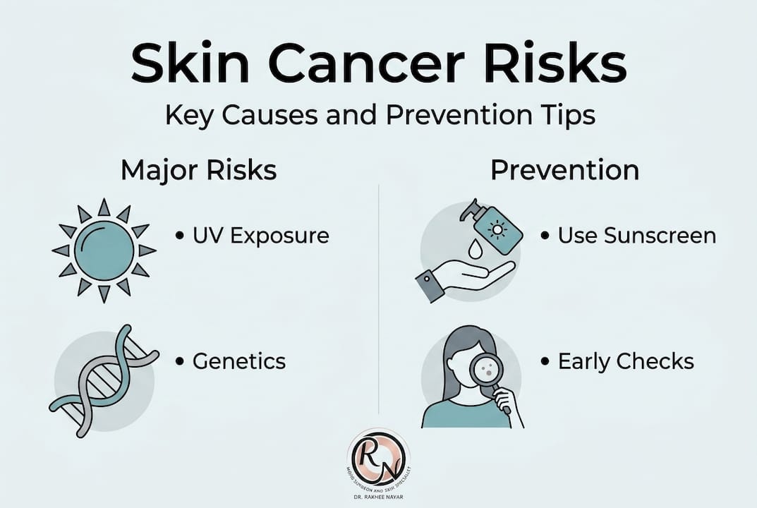
Consistent UV protection, including daily SPF use and avoiding sunbeds, alongside regular professional skin checks, can reduce your risk by up to 90%. Fair-skinned individuals should treat sun protection as a year-round clinical priority, not a summer habit.
What genetic factors make skin cancer more likely?
Mutations in genes such as PTCH1, TP53, and BRAF significantly increase skin cancer risk. Fair skin associated with MC1R variants raises risk substantially, making genetic awareness an important part of your personal prevention strategy.
How does Mohs surgery help with skin cancer treatment?
Mohs surgery removes cancerous tissue layer by layer, examining each layer under a microscope before proceeding. This approach achieves the highest cure rates while sparing as much healthy skin as possible, making it especially valuable on the face.
What are the early signs to watch for in skin cancer?
Look for new lumps, sores that do not heal within four weeks, persistent redness, or any mole that changes in shape, colour, or size. Remember that skin cancers progress invisibly for years, so visible changes are already late-stage signals worth acting on immediately.
Recommended
- Types of skin cancer and advanced facial treatments UK
- High-risk skin cancer: Expert treatment guide for UK patients
- Skin Cancer Detection – Expert Mohs Surgery | UK
- Skin Cancer on the Nose – Symptoms & Treatment – Expert Mohs Surgery | UK
Filed under Skin Cancer Explained