A melanoma mole is defined as a pigmented skin lesion showing malignant change, and it represents the most dangerous form of skin cancer. Melanoma accounts for the majority of skin cancer deaths despite being less common than basal cell carcinoma or squamous cell carcinoma. The ABCDE criteria, recommended by NICE, Cancer Research UK, and the British Association of Dermatologists (BAD), provide the primary framework for identifying suspicious moles. Recognising these signs early is clinically significant: five-year survival approaches 99% when melanoma is caught at its earliest stage. Miss Rakhee Nayar, GMC-registered Consultant Plastic Surgeon and Mohs specialist, advises that any mole showing change warrants prompt clinical assessment.
1. What are the ABCDE signs of a melanoma mole?
The ABCDE criteria are the primary recommended framework for identifying suspicious moles that may be melanoma. Each letter maps to a specific visual feature that clinicians and patients can assess. Understanding all five gives you a structured way to evaluate any mole on your skin.

Asymmetry
A normal mole is roughly symmetrical. If you draw an imaginary line through the centre, both halves should mirror each other. A melanoma mole is often asymmetrical, meaning one half looks noticeably different from the other.
Border irregularity
Benign moles have smooth, well-defined edges. Suspicious lesions tend to have ragged, notched, or blurred borders. Irregular edges suggest the cells are not growing in a controlled pattern.
Colour variation
A uniform brown or tan colour is typical of a benign mole. Melanoma often shows multiple shades within a single lesion, including brown, black, red, white, or blue. Colour variation within one mole is a significant warning sign.
Diameter
Moles larger than 6 millimetres, roughly the size of a pencil eraser, carry higher suspicion for melanoma. That said, some melanomas are diagnosed at smaller sizes, so diameter alone is not a definitive rule.
Evolution
Any change in a mole’s size, shape, colour, or texture over weeks or months is a red flag. Evolution is arguably the most clinically important feature because it signals active cellular change. A mole that bleeds, crusts, or becomes raised without explanation requires urgent evaluation.
Pro Tip: Photograph your moles monthly in consistent lighting. Side-by-side comparison over time makes subtle evolution far easier to detect than relying on memory alone.
These five features work together. No single feature confirms malignancy, and a constellation of signs should prompt clinical evaluation rather than any one criterion in isolation.
2. What additional signs may indicate a suspicious mole?
The ABCDE framework covers the most common melanoma presentations, but several important warning signs fall outside it. Recognising these additional skin cancer signs can be the difference between early and late diagnosis.
The ugly duckling sign
The ugly duckling method asks you to look at your moles as a group rather than individually. Most moles on a given person share a family resemblance in size, shape, and colour. A mole that looks distinctly different from all the others, the “ugly duckling,” warrants clinical attention even if it does not meet strict ABCDE criteria. This approach is particularly useful for patients with many moles.
Bleeding, itching, and tenderness
A mole that bleeds without injury, itches persistently, or feels tender to touch is behaving abnormally. These symptoms suggest the lesion may be growing or ulcerating beneath the surface. They are not always present in melanoma, but their appearance in a changing mole should prompt same-week assessment.
Amelanotic melanoma
Amelanotic melanomas lack the pigment that makes most melanomas visible. They can appear as skin-coloured, pink, or red bumps and are frequently mistaken for benign lesions such as cysts or dermatitis. Between 2% and 8% of melanomas are amelanotic, making them a clinically significant diagnostic challenge. Because they do not look like a typical mole, the ABCDE criteria are less reliable for this subtype.
The key lesson here is that absence of dark pigment does not rule out melanoma. Any new or changing lesion that does not resolve within a few weeks deserves professional assessment, regardless of its colour.
3. What mole types and risk factors increase melanoma risk?
Certain mole types and personal characteristics raise the statistical likelihood of developing melanoma. Understanding your own risk profile helps you decide how frequently to seek skin checks and how urgently to act on changes.
Higher-risk mole types
Atypical (dysplastic) moles are larger than average, have irregular borders, and show uneven colour. They are not cancerous themselves, but they signal elevated risk. Individuals with 10 or more atypical moles are 12 times more likely to develop melanoma than those without them. That figure underscores why regular dermatological surveillance matters for this group.
Congenital moles are present at birth. Large congenital moles, particularly those exceeding 20 centimetres in diameter, carry a measurably higher lifetime risk of malignant transformation than moles acquired in adulthood.
Numerous common moles also increase risk. Having more than 50 ordinary moles is itself a recognised risk factor, independent of whether any individual mole appears atypical.
Personal risk factors
The following factors are associated with increased melanoma risk:
- Pale skin, light hair, or light eyes. Reduced melanin provides less natural protection against ultraviolet radiation.
- Family history of melanoma. A first-degree relative with melanoma roughly doubles your personal risk.
- Previous melanoma diagnosis. Patients who have had one melanoma are at significantly higher risk of a second primary tumour.
- Immunosuppression. Patients on long-term immunosuppressive therapy, including organ transplant recipients, face elevated risk.
- Extensive sun exposure or sunbed use. Cumulative ultraviolet exposure and a history of sunburn, particularly in childhood, are established risk factors.
- Age. Melanoma risk increases with age, though it is one of the more common cancers in adults under 50.
| Risk factor | Clinical significance |
|---|---|
| 10+ atypical moles | 12 times higher melanoma risk |
| Family history (first-degree) | Approximately double the baseline risk |
| Large congenital mole (>20 cm) | Elevated lifetime malignant transformation risk |
| Previous melanoma | Significantly raised risk of second primary |
| Immunosuppression | Reduced immune surveillance of abnormal cells |
Approximately one in three melanomas develop within a pre-existing mole; the remainder arise on previously normal skin. This means that monitoring existing moles is necessary but not sufficient. Full skin checks, including areas rarely exposed to the sun, remain important.
4. How is a suspicious mole diagnosed and treated?
Clinical diagnosis and treatment follow a defined pathway. Knowing what to expect reduces anxiety and helps you engage more effectively with your clinical team.
Dermoscopy
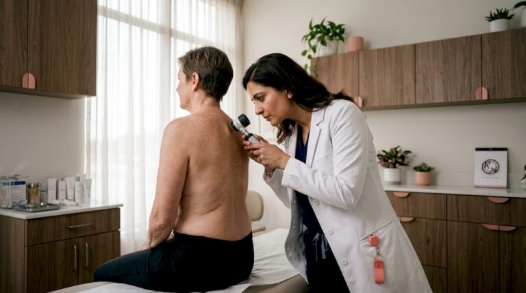
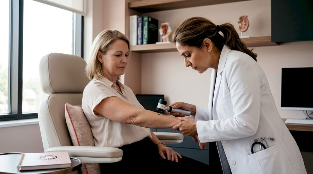
Dermoscopy is a non-invasive technique in which a specialist uses a handheld device with magnification and polarised light to examine structures beneath the skin surface. It allows clinicians to assess pigment patterns, vascular structures, and other features invisible to the naked eye. Dermoscopy significantly improves diagnostic accuracy compared with visual inspection alone.
Biopsy
A biopsy is the definitive diagnostic step. The suspicious lesion is excised or sampled and sent for histopathological analysis. The pathology report confirms whether melanoma is present, identifies the subtype, and measures the Breslow thickness, which is the depth of invasion. Breslow thickness is the single most important prognostic factor in early melanoma.
Wide local excision
Wide local excision (WLE) is the standard surgical treatment for confirmed melanoma. The surgeon removes the tumour along with a margin of surrounding healthy tissue. The margin width is determined by Breslow thickness and follows NICE guidelines. WLE is effective for most melanomas on the trunk and limbs.
Mohs micrographic surgery
Mohs micrographic surgery excises cancerous tissue layer by layer, with microscopic margin assessment at each stage until all margins are clear. This approach preserves the maximum amount of healthy tissue while achieving complete tumour removal. It is particularly valuable for melanomas on the face and other cosmetically sensitive areas where tissue conservation matters. Miss Rakhee Nayar is dual-trained in both Mohs surgery and plastic surgery, which means reconstruction and excision are planned together from the outset.
Pro Tip: Ask your surgeon specifically about Breslow thickness after your biopsy result. This single measurement guides the recommended excision margin and informs your staging, so understanding it helps you ask the right questions at your next appointment.
For a detailed comparison of surgical approaches, the Mohs vs standard excision guide from Rakhee Nayar – Mohs Surgeon and Skin Specialist sets out the clinical criteria that determine which procedure is appropriate.
The early detection benefits for melanoma are well established. Five-year survival approaches 99% for stage I disease. That figure drops substantially with each increase in stage, which is why acting on a changing mole promptly rather than waiting is the clinically correct decision.
Key takeaways
Early recognition of a melanoma mole using the ABCDE criteria, combined with prompt specialist assessment, gives patients the best chance of successful treatment and high survival rates.
| Point | Details |
|---|---|
| ABCDE criteria are the primary tool | Asymmetry, Border, Colour, Diameter over 6 mm, and Evolution guide self-assessment. |
| Ugly duckling sign adds detection value | A mole that looks different from your others warrants clinical review even without ABCDE features. |
| Atypical moles multiply risk significantly | Ten or more atypical moles raises melanoma risk 12 times compared with the general population. |
| Amelanotic melanoma is easily missed | Pigment-free melanomas appear pink or skin-coloured and do not respond to standard ABCDE checks. |
| Early detection transforms outcomes | Five-year survival approaches 99% when melanoma is caught at its earliest stage. |
Miss Nayar’s perspective: what I see that patients often miss
Patients frequently arrive at consultation having monitored a mole for months, reassuring themselves that it “hasn’t changed much.” The problem is that subtle evolution is genuinely difficult to detect without a baseline photograph and consistent lighting. I see this pattern repeatedly, and it is the single most preventable delay in melanoma diagnosis.
The ABCDE framework is sound, but it was designed as a clinical teaching tool, not a definitive self-diagnosis checklist. A mole can satisfy only one or two criteria and still be melanoma. Conversely, a mole with several atypical features can prove benign on biopsy. What matters is the clinical context: your skin type, your mole count, your family history, and whether anything has changed. No app or photograph replaces that assessment.
The patients I am most concerned about are those who have amelanotic lesions. These look nothing like the classic dark, irregular mole. They present as a persistent pink or red spot that does not heal, and they are routinely dismissed as eczema or a minor skin irritation. If you have a lesion that has been present for more than six weeks without resolving, please seek a specialist opinion regardless of its colour.
Mohs surgery, when indicated, gives me the ability to confirm clear margins in real time during the procedure. For melanomas on the face or ears, that matters enormously for both cure and cosmetic outcome. The reconstruction is planned before the first incision, not as an afterthought. That dual-trained approach is what distinguishes specialist Mohs care from a standard excision performed in isolation.
My advice is simple. Photograph your skin regularly. Know your risk factors. Act on change, not on certainty.
— Miss Rakhee Nayar
Specialist skin cancer care at Mohs Surgeon UK
Rakhee Nayar – Mohs Surgeon and Skin Specialist offers consultant-led assessment and treatment for patients with suspicious moles and confirmed melanoma at Circle Cheshire in North West England.

Miss Nayar’s dual training in plastic surgery and Mohs micrographic surgery means that excision and reconstruction are planned together, with clear margins confirmed before the wound is closed. Private consultations and e-consultations are available for UK and international patients. For patients who have received a melanoma diagnosis or are concerned about a changing mole, the melanoma treatment guide and the Mohs surgery service page set out the full range of treatment pathways available. To book a consultation, visit mohssurgeon.co.uk.
This article is for informational purposes only and does not constitute medical advice. Consult a GMC-registered specialist for assessment and treatment of any suspicious skin lesion.
FAQ
What does a melanoma mole look like?
A melanoma mole typically shows asymmetry, irregular borders, multiple colours, a diameter greater than 6 mm, or visible change over time. Some melanomas, known as amelanotic melanomas, appear pink or skin-coloured and lack typical dark pigmentation.
How quickly does melanoma develop in a mole?
Melanoma can develop over weeks to months, which is why any mole showing change within a short period warrants prompt clinical review. Evolution, meaning any change in size, shape, colour, or texture, is one of the most important warning signs.
Can a normal-looking mole be melanoma?
Yes. Amelanotic melanomas in particular can appear as unremarkable pink or skin-coloured bumps. The ugly duckling sign helps identify lesions that look different from your other moles, even when they do not meet standard ABCDE criteria.
What is the treatment for a melanoma mole?
Treatment depends on the stage and location. Wide local excision is standard for most melanomas, while Mohs micrographic surgery is used for lesions on the face or cosmetically sensitive areas where tissue preservation is a priority.
When should I see a specialist about a mole?
See a GMC-registered specialist if a mole changes in size, shape, or colour, bleeds without injury, itches persistently, or simply looks different from your other moles. Early assessment is the most effective way to improve outcomes.