Key Takeaways:
- Mohs surgery uses its own on-site lab so tissue can be frozen, cut, stained, and read while you wait, instead of being sent away for days.
- A cryostat freezes each sample in seconds, and a microtome inside it slices sections a few micrometres thick, thinner than a human hair.
- Every piece of tissue is colour-coded on a hand-drawn map before it’s processed, so any remaining cancer can be traced back to an exact spot on your skin.
- A biomedical scientist runs the lab equipment, but it’s the Mohs surgeon, not a separate pathologist, who reads every slide in person.
Patients see the surgery. What they rarely see is the small lab next door where the actual decision gets made, whether the cancer is gone or whether another layer needs to come out. It’s a handful of pieces of equipment and one highly trained person, working together in the time it takes you to sit in the waiting room.
Here’s what’s actually happening behind that door, and why it’s set up so differently from a standard hospital lab.
The Mohs lab at a glance
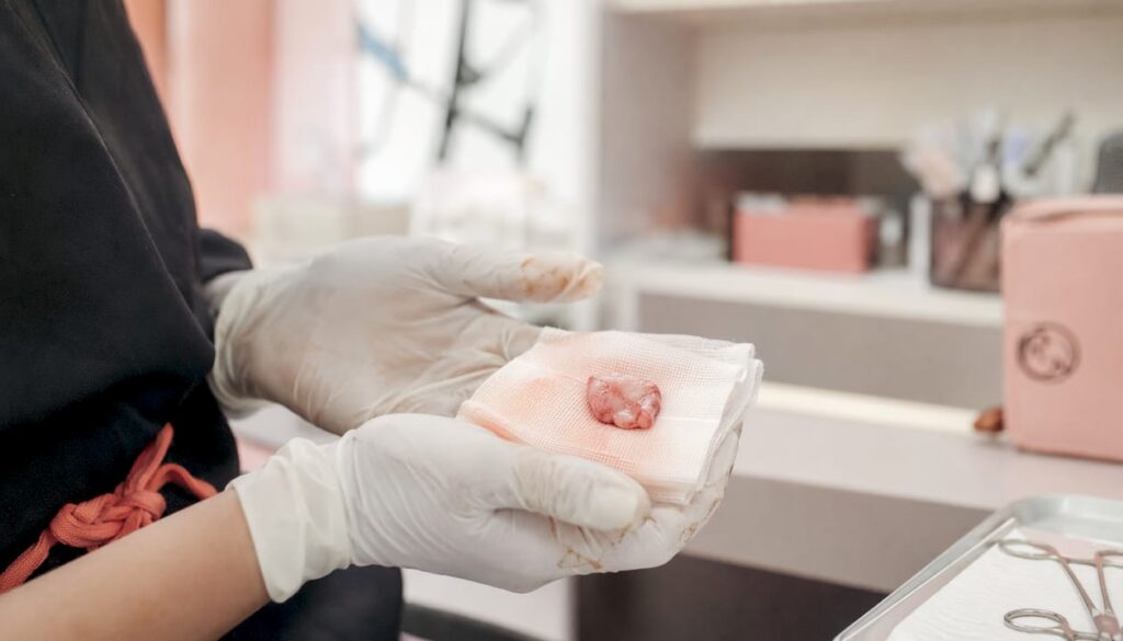
| Equipment | What it does |
|---|---|
| Cryostat | Freezes each tissue sample solid in seconds, ready to be sliced. |
| Microtome | Built into the cryostat, cuts sections a few micrometres thick. |
| Mapping board | A colour-coded diagram linking each slice back to an exact spot on your skin. |
| Staining rack | Applies dye so the cell structures show up clearly under the microscope. |
| Microscope | Where the surgeon examines the entire margin in person. |
Why Mohs surgery needs its own on-site lab
A standard skin biopsy is usually sent away to an off-site pathology lab, processed over several days, and the result comes back by letter or phone call.
Mohs surgery can’t work that way. Because each stage depends on the result of the one before it, the lab has to sit next door to the operating room, staffed and ready, so a sample can go from tissue to answer in minutes rather than days. What that wait actually feels like from the patient’s side is covered on the what happens between Mohs surgery stages page.
The cryostat, freezing tissue in seconds
The cryostat is a freezing chamber built around a cooled metal plate. The tissue is mounted in a gel-like freezing medium and cooled to roughly minus 20 to minus 22 degrees Celsius for most skin, slightly colder for fatty tissue and slightly warmer for cartilage, which shatters if it gets too cold.
Speed matters here. According to the National Institutes of Health’s clinical reference on Mohs tissue processing, freezing too slowly lets ice crystals form inside the cells, which tears holes in the tissue and makes it unreadable under the microscope. A well-run cryostat avoids that entirely.
The microtome, cutting sections thinner than a human hair
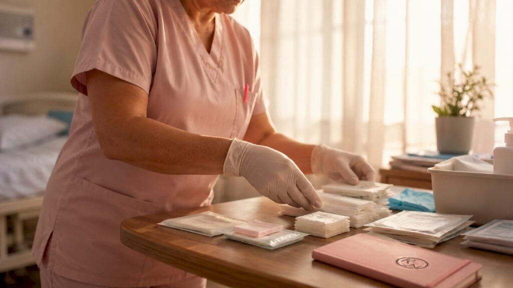
Built into the cryostat is a microtome, a blade fine enough to shave off sections roughly 5 to 6 micrometres thick. A human hair is around 70 micrometres across, so each slice is a small fraction of that.
At that thickness the frozen tissue is extremely fragile. It’s traditionally lifted and flattened onto the glass slide with a fine brush, by hand, before it has any chance to curl or tear.
The mapping board, colour-coding where each piece came from
Before any freezing or cutting happens, the removed tissue is divided into pieces and each one is marked with a different coloured dye, then sketched onto a diagram of your wound.
That map is what makes Mohs surgery precise rather than just thorough. If cancer shows up on one slide, the colour tells the surgeon exactly which piece it came from, and the diagram tells them exactly where on your skin that piece sat, down to the position on the clock face. How that map is built is covered in more depth by skin cancer mapping.
Staining the slides so the surgeon can see what’s there
A freshly cut section is colourless and almost impossible to read under a microscope. Staining fixes that. The most common method is haematoxylin and eosin, usually shortened to H&E, run through roughly ten steps in quick succession.
Haematoxylin stains the cell nuclei a blue-purple, eosin counterstains the surrounding structures pink, and the slide is then dehydrated, cleared, and sealed under a coverslip. What comes out the other end is a slide where cancerous cells stand out clearly against healthy tissue.
The biomedical scientist, the specialist you rarely meet
Every step from freezing to staining is run by a trained laboratory specialist, called a biomedical scientist in the UK, registered with the Health and Care Professions Council in the same way a pharmacist or physiotherapist is.
You won’t usually meet them, but they’re working the whole time you’re in the waiting room. Getting one slide from raw tissue to a readable stain is a skilled, hands-on process, and a poorly cut or poorly stained section can hide the very thing the surgeon is looking for.
The microscope, where the surgeon reads your margin in person
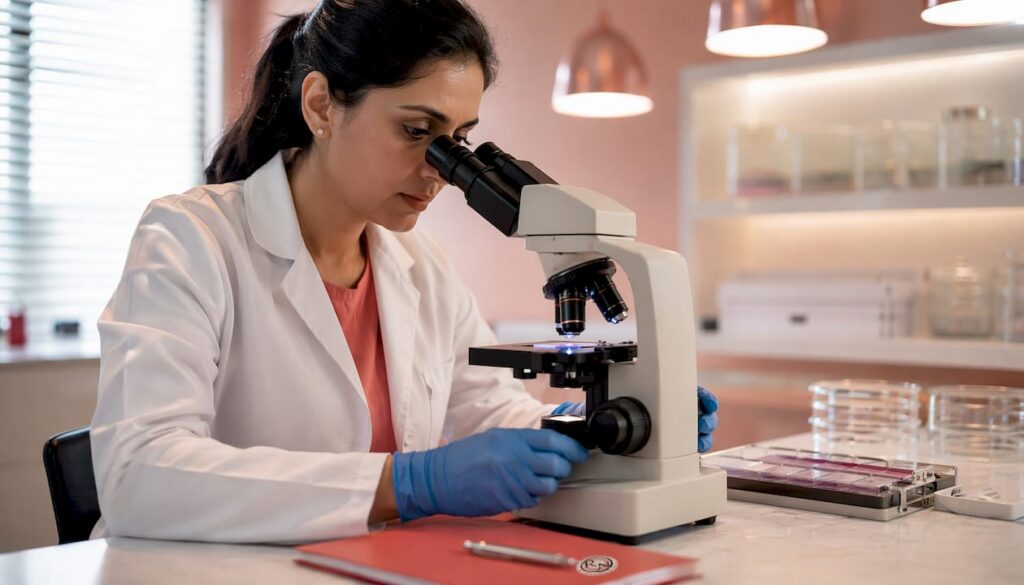
This is the step that sets Mohs surgery apart from a standard biopsy. The slide isn’t sent to a separate pathologist to interpret days later. Your own surgeon sits down at the microscope and reads it themselves, checking the entire cut edge and undersurface of every piece against the colour-coded map.
If every margin is clear, you’re done. If not, the surgeon already knows the precise location to remove another, usually much smaller, layer, because they read the map themselves rather than relying on someone else’s written report.
Why this differs from a standard biopsy lab
A standard pathology lab is built for volume, processing samples from many different clinics and hospitals, embedded in wax rather than frozen, which takes days to complete properly.
A Mohs lab is built for one thing: speed without losing accuracy, for a single patient’s tissue, on a single day. How long Mohs surgery actually takes covers how that lab turnaround fits into your overall appointment.
Curious to see how it all fits together
Knowing what’s happening in the lab tends to make the waiting easier, it isn’t a black box, it’s a short, well-rehearsed process with a specific piece of equipment for every step. The surgical side that this lab work supports is covered separately in what Mohs micrographic surgery actually involves.
If you have questions about your own case before booking, get in touch through the contact page to arrange a consultation with Miss Rakhee Nayar.
FAQ
Does every Mohs surgery clinic have its own lab?
Any clinic offering true Mohs surgery needs same-day access to a lab set up for frozen sectioning. Without it, results can’t come back while you wait, which is the whole point of the technique.
Who reads the slides, a pathologist or the surgeon?
The Mohs surgeon reads every slide themselves, in person, rather than sending it to a separate pathologist. That’s one of the main things that separates Mohs surgery from a standard biopsy.
Why is the tissue frozen instead of processed the normal way?
Standard tissue processing uses wax embedding, which takes days to complete properly. Freezing the tissue in a cryostat lets it be sliced and stained within minutes, which is what makes same-day results possible.
What is a biomedical scientist’s role in Mohs surgery?
They run the cryostat, cut the frozen sections, and carry out the staining, working closely with the surgeon throughout the appointment. In the UK, they’re registered with the Health and Care Professions Council.
What is the coloured map used for?
It links each piece of tissue on the slide back to an exact location on your skin. If cancer is found on one piece, the map tells the surgeon precisely where to remove more tissue from, rather than guessing.
Recommended
- What Happens Between Mohs Surgery Stages? Understanding the Interval
- Understanding skin cancer mapping for precise treatment
- How Long Does Mohs Surgery Actually Take? A Timeline From Consultation to Closure
- What is Mohs Micrographic Surgery? | Miss Rakhee Nayar
Filed under Mohs Surgery