Key takeaways
- Bowen’s disease is squamous cell carcinoma in situ, meaning the abnormal cells are confined to the outermost layer of skin and have not invaded deeper tissue
- It usually appears as a slow-growing, persistent, scaly or crusted red patch, classically on the lower legs
- Left untreated, a minority of cases progress to invasive squamous cell carcinoma over time
- It is treated in several ways depending on size and location, and is highly treatable when caught at this stage
Bowen’s disease is a name that understandably worries people when they first hear it attached to their own skin. It sounds separate from skin cancer, but it is in fact an early, contained form of it. Understanding exactly what that means, and what it does not mean, is the quickest way to put it in perspective.
What is Bowen’s disease
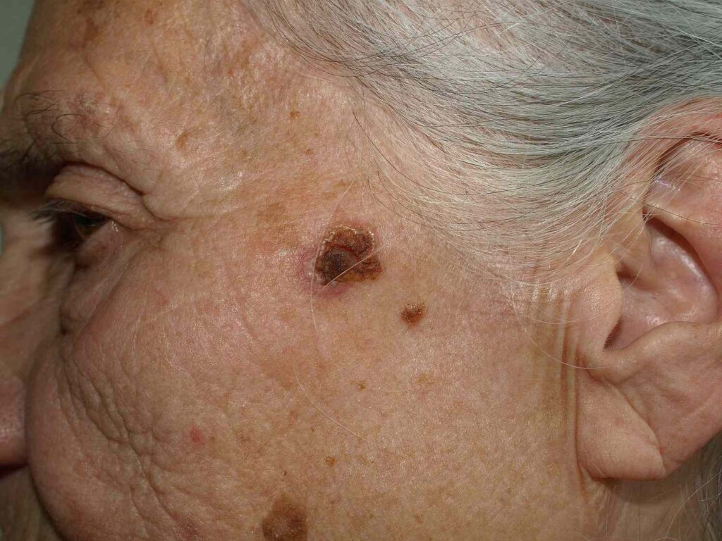
Bowen’s disease is squamous cell carcinoma in situ. That means the abnormal, cancerous-looking cells are present, but they are confined entirely to the epidermis, the outermost layer of skin, and have not broken through into the dermis beneath. It is named after the American dermatologist John T. Bowen, who first described it in 1912.
Why it’s also called SCC in situ
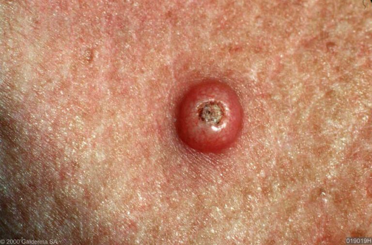
“In situ” is Latin for “in place.” It is used across several types of cancer, including melanoma in situ, to describe a stage where abnormal cells exist but have not yet gained the ability to invade surrounding tissue or spread elsewhere. For squamous cell carcinoma, Bowen’s disease is that in situ stage. It is sometimes thought of as a halfway point between actinic keratosis and fully invasive squamous cell carcinoma, though not every case follows a neat line between the two.
What it looks like
Typical appearance
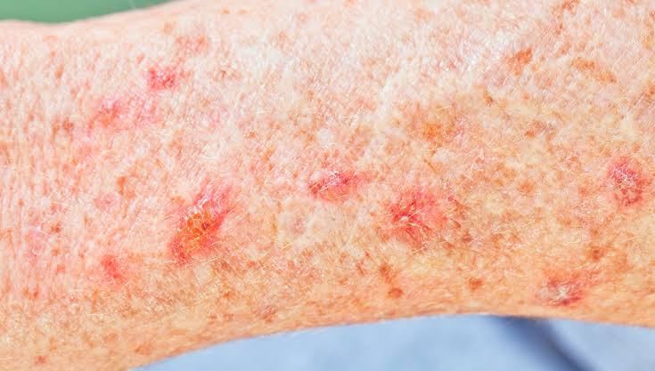
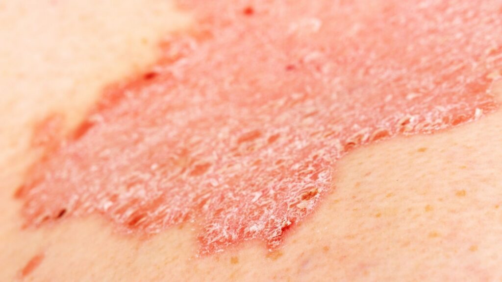
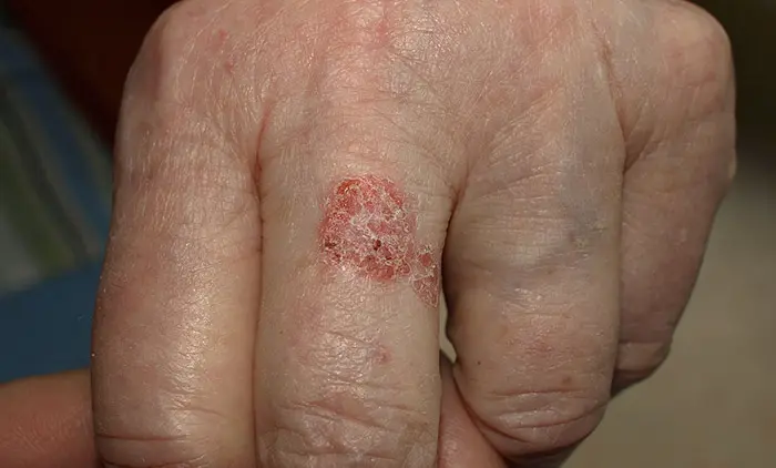
A typical patch is red or pink, scaly, and well-defined, often described as looking a little like a persistent patch of eczema or psoriasis that has not responded to the usual creams. It tends to grow very slowly, sometimes over years, and can become slightly raised or crusted as it develops. It is usually larger than a typical actinic keratosis, often a centimetre or more across, and solitary rather than appearing in clusters.
Where it tends to develop
Bowen’s disease classically appears on the lower legs, particularly in women, though the reasons for this pattern are not fully settled. It can also occur on the hands, scalp, face, and other sun-exposed skin. A separate condition that is sometimes confused with it by name alone is genital Bowen’s disease, which affects the genital skin and is caused by HPV infection rather than sun exposure. The two share a name and a similar cell appearance under the microscope, but they are different conditions with different causes, and this guide is focused on the sun-related, non-genital form.
What causes it
Cumulative ultraviolet exposure over many years is the main driver of non-genital Bowen’s disease, in the same way it drives actinic keratosis and squamous cell carcinoma more broadly. Fair skin, a long history of outdoor sun exposure, and in some cases a weakened immune system or prior arsenic exposure (historically relevant, now rare) are recognised risk factors.
Why it is taken seriously despite being “in situ”
Because the abnormal cells have not invaded deeper tissue, Bowen’s disease carries a much lower risk than invasive squamous cell carcinoma. However, a minority of untreated cases do eventually progress to invasive disease over months to years, particularly if left completely unmanaged. That risk, combined with how reliably treatable it is at this stage, is why specialists recommend treating it rather than simply monitoring it.
How it’s diagnosed
A persistent, slow-growing scaly patch that has not responded to standard eczema or psoriasis treatment is usually assessed with a skin biopsy, which confirms the diagnosis under the microscope and rules out an invasive component. You can read more about what to expect from a skin cancer biopsy if this has been suggested.
How Bowen’s disease is treated
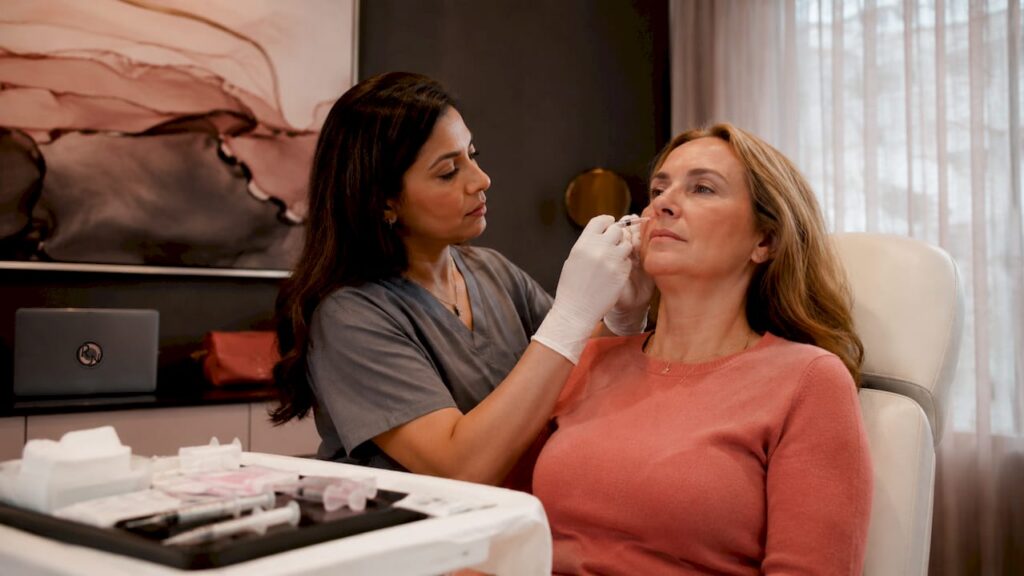
Treatment is chosen based on the size, location, and number of patches, and the person’s general health. Common options include:
| Treatment | Typical use |
|---|---|
| Cryotherapy | Smaller, well-defined patches |
| Topical chemotherapy or immune cream | Larger or multiple patches, particularly on the lower leg |
| Photodynamic therapy | Larger patches, or areas where scarring is a concern |
| Curettage and cautery | Well-defined patches where tissue sampling is also useful |
| Surgical excision | Patches where complete removal and margin control matter most |
Lower leg skin in particular can be slow to heal, which is a factor your specialist will weigh when recommending the most suitable option for your situation.
Outlook and follow-up
With appropriate treatment, the outlook for Bowen’s disease is very good. Because it reflects a broader pattern of sun-damaged skin, follow-up checks are usually recommended to catch any new patches or, rarely, recurrence at the treated site. Our guide to spotting the early signs of skin cancer is a useful reference for what else to watch for in the meantime.
FAQ
Is Bowen’s disease actually cancer?
Yes, technically. It is squamous cell carcinoma in situ, meaning cancerous cells are present but confined to the outer layer of skin. It is not invasive, which is why the outlook is generally very good.
Will Bowen’s disease definitely turn into invasive cancer if untreated?
No. Most cases remain in situ indefinitely, but a minority do progress to invasive squamous cell carcinoma over time, which is why treatment is generally recommended rather than a watch-and-wait approach.
Is Bowen’s disease on the genitals the same condition?
No. Genital Bowen’s disease looks similar under the microscope but is caused by HPV rather than sun exposure, and is managed differently. Always mention the exact location when seeking advice.
Does Bowen’s disease leave a scar after treatment?
It depends on the treatment used and the size of the patch. Your specialist will discuss the likely cosmetic outcome of each option before you decide.
Recommended next step
If you have a persistent scaly patch that has not responded to standard treatment, it is worth having it properly assessed. Learn more about squamous cell carcinoma and its early forms, or book an assessment with Miss Rakhee Nayar.
This article is for general information and does not replace a face-to-face medical assessment. If you are concerned about a specific mark on your skin, please seek an in-person review.
Filed under Skin Cancer Explained