Key takeaways
- A keratoacanthoma is a fast-growing skin lump that can look and behave almost identically to squamous cell carcinoma
- Even under the microscope, the two can be genuinely difficult to tell apart
- Because of this, most specialists treat a keratoacanthoma as if it were cancer, by removing it
Few skin lumps appear as suddenly or grow as quickly as a keratoacanthoma. A smooth, dome-shaped bump can go from barely noticeable to a centimetre or more across in a matter of weeks, which understandably alarms most people who notice one. The frustrating truth is that this same rapid growth is also a feature of squamous cell carcinoma, and the two conditions can look near-identical, even to specialists examining a biopsy down a microscope.
What a keratoacanthoma actually is
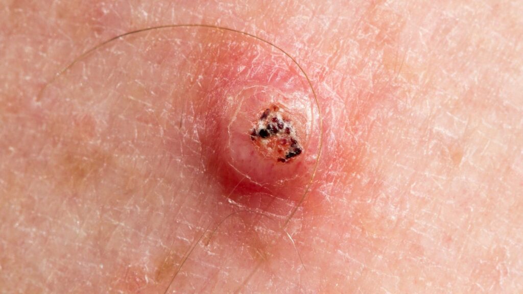
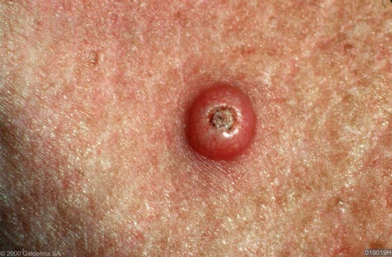
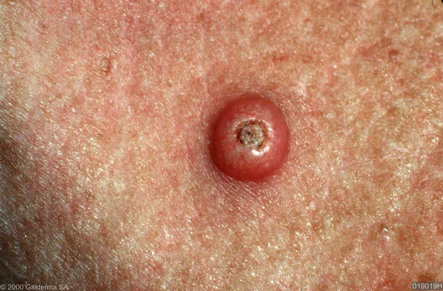
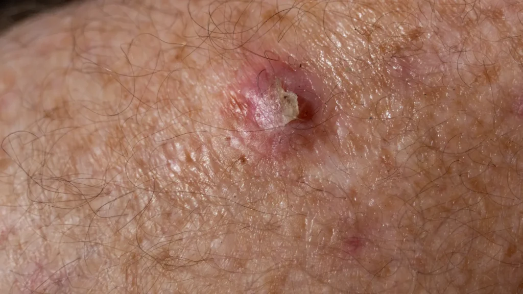
A keratoacanthoma is a skin growth that develops from hair follicle cells, typically appearing as a firm, dome-shaped nodule with a central crater filled with keratin (the same protein that makes up skin, hair, and nails). It usually appears on sun-exposed skin, such as the face, ears, forearms, and backs of the hands, and is most common in older adults with a long history of sun exposure.
Why it’s so often mistaken for squamous cell carcinoma
Keratoacanthoma and well-differentiated squamous cell carcinoma share the same rapid growth pattern, a similar dome shape, and, critically, a very similar cell structure when examined under a microscope. Some pathologists consider keratoacanthoma to sit on a spectrum with squamous cell carcinoma rather than being a wholly separate entity. This overlap is not a gap in diagnostic skill. It is a genuine, well-recognised grey area in dermatopathology.
The signs that help tell them apart
- Speed of onset. A keratoacanthoma typically appears and grows to its full size within 4 to 8 weeks, faster than most squamous cell carcinomas develop.
- The crater. A central, keratin-filled crater with a volcano-like appearance is more characteristic of keratoacanthoma, though it is not exclusive to it.
- What happens next. Left alone, some keratoacanthomas plateau and then slowly shrink and regress over several months, something true squamous cell carcinoma essentially never does.
- Microscopic detail. A pathologist looks for specific architectural features and the overall growth pattern, though as noted above, this does not always give a clean answer.
- Size and depth on removal. The full picture often only becomes clear once the whole lesion has been removed and examined in its entirety, rather than from a small sample.
How keratoacanthoma develops: rapid growth, then possible regression
The hallmark feature of keratoacanthoma is its three-phase course: a rapid growth phase over weeks, a plateau, and then, in some cases, spontaneous regression over several months, sometimes leaving a small scar behind. This pattern is well documented and is one of the few reliable biological differences from squamous cell carcinoma, which does not spontaneously shrink on its own. The difficulty is that nobody can predict in advance, from looking at a growing lump in its early weeks, whether it will follow this regressive course or whether it is actually an invasive cancer that will keep growing.
Why some specialists treat every keratoacanthoma as if it were cancer anyway
Given the overlap described above, waiting to see whether a lump regresses on its own is not considered an acceptable strategy in most cases. The standard, cautious approach is to treat a keratoacanthoma as if it were squamous cell carcinoma until proven otherwise, which in practice means surgical removal. This avoids the risk of leaving a genuine cancer to grow unchecked while waiting for a spontaneous regression that may never come.
How it’s diagnosed and treated
A rapidly growing dome-shaped lump is usually assessed with a biopsy or, more often, complete surgical excision, since examining the whole lesion gives the clearest picture. Our guide on preparing for a skin cancer biopsy explains what that process involves. Surgical removal, sometimes using Mohs micrographic surgery for lesions in cosmetically or functionally sensitive areas, both treats the lesion and provides a definitive diagnosis.
A word on self-diagnosis

Because even trained specialists can struggle to separate keratoacanthoma from squamous cell carcinoma by appearance alone, trying to reach a confident verdict from a photo or a mirror is not realistic. The practical takeaway is simpler than the biology: any new lump that grows quickly over a few weeks deserves a proper in-person assessment, regardless of which of the two conditions it ultimately turns out to be. Our broader guide to spotting the early signs of skin cancer covers the warning signs worth acting on.
FAQ
Can a keratoacanthoma just be left to go away on its own?
In theory some do regress without treatment, but because it cannot be reliably distinguished from squamous cell carcinoma in advance, leaving it untreated is not generally recommended. Removal is the safer course of action.
Is a keratoacanthoma dangerous?
On its own, a true keratoacanthoma is considered low risk and does not spread. The danger lies in the diagnostic uncertainty, since an actual squamous cell carcinoma can look identical at first.
How fast does a keratoacanthoma grow?
Most reach their full size within 4 to 8 weeks, which is notably faster than typical squamous cell carcinoma growth, though this alone is not enough to confirm the diagnosis.
Recommended next step
If you have noticed a lump that has grown quickly over the past few weeks, it is worth having it assessed promptly rather than waiting to see what it does. Book a skin cancer assessment with Miss Rakhee Nayar for an accurate diagnosis and, where needed, prompt treatment.
This article is for general information and does not replace a face-to-face medical assessment. If you are concerned about a specific lump or mark on your skin, please seek an in-person review.
Filed under Basal and Squamous Cell Carcinoma