

Cysts on the head are benign, fluid-filled or keratin-containing lumps that form beneath the scalp skin, most commonly arising from hair follicles or epidermal cells. Pilar cysts account for approximately 90% of scalp cysts, making them by far the most frequently encountered type in clinical practice. The second most common type is the epidermoid cyst, which forms from epidermal skin cells rather than follicle tissue. Both types are almost always benign, but a new or changing lump on the scalp always warrants professional assessment to rule out more serious conditions. This article provides clinically accurate guidance on causes, symptoms, treatment, and when to seek specialist advice, directed by Miss Rakhee Nayar, Consultant Plastic Surgeon and Mohs specialist.
What causes cysts on the head and how do they develop?
Scalp cysts form through two distinct biological processes, depending on their type. Understanding the difference helps clarify why some patients develop multiple cysts while others have only one.
Pilar (trichilemmal) cysts
Pilar cysts originate from the outer root sheath cells of the hair follicle. When these cells accumulate keratin, a fibrous structural protein, they form a sac beneath the scalp surface. The cyst wall is derived from follicular epithelium, which gives pilar cysts their characteristic thick, firm shell.
A strong hereditary pattern is well established in pilar cysts. Family history is present in about 50% of cases, and the inheritance follows an autosomal dominant pattern. This means a patient with a parent who has pilar cysts has a roughly one in two chance of developing them too. Multiple cysts occur in 70% of patients with this type, so finding two or three lumps on the scalp at once is entirely consistent with a benign hereditary condition.
Epidermoid cysts
Epidermoid cysts arise from epidermal skin cells that become trapped beneath the skin surface. This can happen after a minor injury, a blocked follicle, or spontaneously. The cyst fills with keratin debris and dead skin cells rather than the compact keratin seen in pilar cysts.
Epidermoid cysts are far less common on the scalp than elsewhere on the body. They tend to be solitary and are not strongly linked to family history. A visible central punctum, a small dark dot on the skin surface, is often present and helps distinguish them clinically from pilar cysts.
- Pilar cysts form from hair follicle outer root sheath cells and contain compact keratin.
- Epidermoid cysts form from trapped epidermal cells and contain softer, cheesy keratin debris.
- Blocked follicles contribute to both types by preventing normal cell shedding.
- Hereditary factors are significant for pilar cysts but less so for epidermoid cysts.
- Location matters: pilar cysts are almost exclusively scalp-based, while epidermoid cysts appear across the body.
Pro Tip: If you have a parent or sibling with multiple scalp lumps, tell your clinician. A family history of pilar cysts changes the clinical picture and may mean a lower threshold for surgical removal is appropriate.
What are the symptoms of scalp cysts, and when should you seek help?
Most scalp cysts cause no symptoms at all. Patients often discover them by chance when washing or brushing their hair. Knowing which features are reassuring and which require prompt attention is the most practical knowledge you can have.
Typical features of a benign scalp cyst
A benign scalp cyst is usually firm and smooth to the touch. It moves freely under the skin when pressed, which is a key sign that it is not attached to deeper structures. Most are painless and grow very slowly over months or years. Size varies from a few millimetres to several centimetres, though most remain under two centimetres.
A lump that moves freely beneath the scalp and has been stable for months is very likely benign. A lump that is fixed, growing rapidly, or associated with skin changes requires professional evaluation without delay.
Warning signs that need medical attention
Signs indicating urgent evaluation include rapid enlargement, persistent pain, redness, warmth, and discharge. These features suggest infection or inflammation within the cyst, a condition called an infected or inflamed cyst. Fever is a rare but serious sign that infection has spread beyond the cyst wall.
The following features should prompt a GP or specialist referral:
- Rapid increase in size over days or weeks
- Redness, warmth, or swelling of the surrounding skin
- Discharge of pus or foul-smelling fluid
- A lump that feels fixed or attached to the underlying scalp
- Persistent pain or tenderness without obvious cause
- Any change in the overlying skin, such as ulceration or irregular pigmentation
Pro Tip: Never attempt to pop or squeeze a scalp cyst. Squeezing a cyst does not remove the sac and risks introducing bacteria, causing a painful infection that complicates subsequent surgical removal.
Self-diagnosis using internet images is unreliable. Professional clinical examination is the only reliable method to distinguish a benign scalp cyst from a lipoma, an abscess, or a skin cancer nodule. If you are uncertain about any lump on your scalp, a consultation with a GMC-registered specialist is the appropriate next step. Patients with a history of skin cancer in the scalp should have any new lump assessed promptly.
Pilar cysts vs epidermoid cysts: what is the difference?
The two main types of scalp cyst are clinically distinct. Recognising their differences helps both patients and clinicians choose the right management approach.
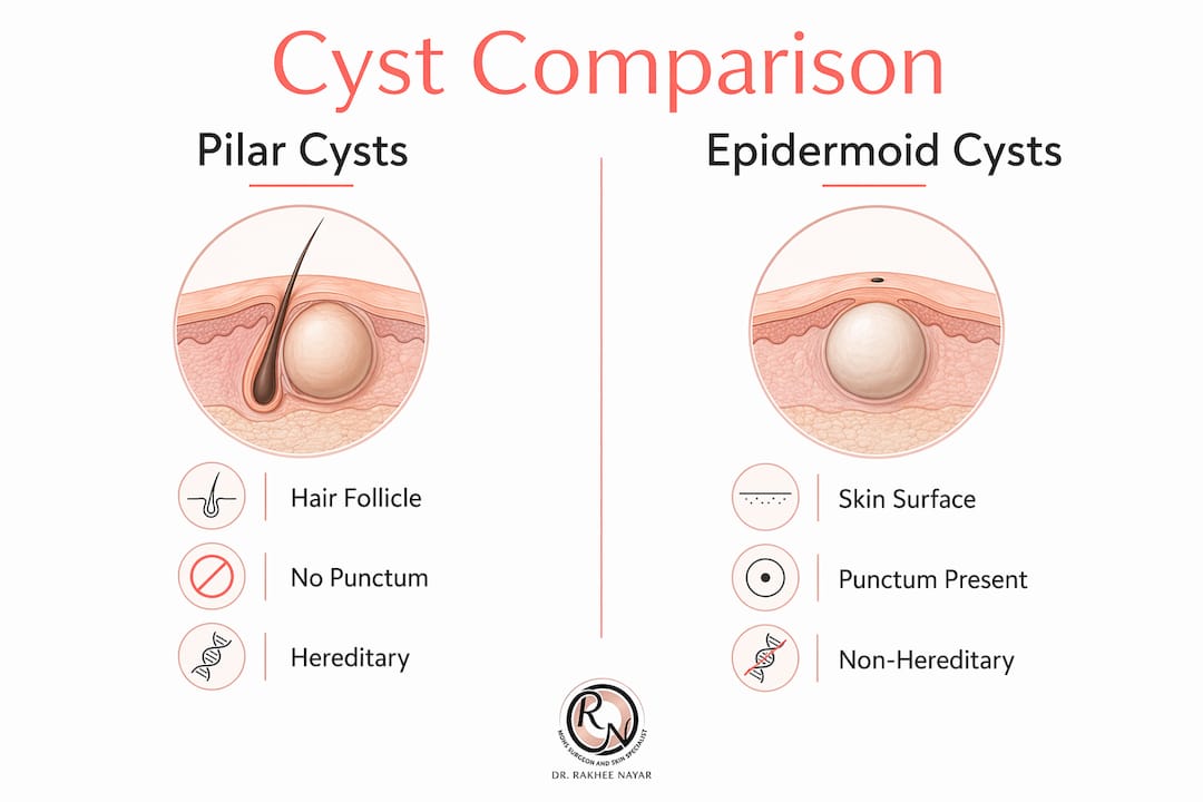
Clinical dermatologists differentiate pilar and epidermoid cysts based on location, presence of a punctum, keratin type, multiplicity, and genetic inheritance patterns. The table below summarises the key features side by side.
| Feature | Pilar (trichilemmal) cyst | Epidermoid cyst |
|---|---|---|
| Cellular origin | Hair follicle outer root sheath | Trapped epidermal skin cells |
| Typical location | Scalp (almost exclusively) | Scalp, face, trunk, genitals |
| Central punctum | Absent | Usually present |
| Contents | Compact, dry keratin | Soft, cheesy keratin debris |
| Proportion of scalp cysts | Approximately 90% | Approximately 10% |
| Multiplicity | Common (70% of patients have multiple) | Usually solitary |
| Hereditary pattern | Autosomal dominant in many cases | Not strongly hereditary |
| Cyst wall | Thick, firm | Thinner, more fragile |
The absence of a punctum in pilar cysts is a clinically useful distinguishing feature. When a clinician examines a firm, mobile scalp lump with no visible punctum and the patient reports a family history of similar lumps, a pilar cyst is the most likely diagnosis. An epidermoid cyst on the scalp, by contrast, often has a visible punctum and is more likely to be solitary.
Both types are benign. Neither type transforms into skin cancer, though any lump that changes character should be reassessed. The distinction matters primarily because it informs surgical planning and guides conversations about recurrence risk and family screening.
What treatment options exist for cysts on the head?
Most scalp cysts do not require treatment if they are stable, painless, and not causing cosmetic concern. The decision to remove a cyst depends on symptoms, size, location, and patient preference.

Conservative management
Watchful waiting is appropriate for small, asymptomatic cysts that have been stable for some time. Patients should monitor for the warning signs described above and return for reassessment if anything changes. No cream, supplement, or home remedy dissolves a cyst sac.
Surgical excision
Surgical excision is the definitive treatment for scalp cysts. The procedure removes the entire cyst, including its wall, under local anaesthetic. Complete removal of the cyst wall is critical to prevent recurrence. Drainage alone, which releases the contents without removing the sac, leads to refilling and recurrence in the majority of cases.
The standard procedure follows these steps:
- Local anaesthetic injection into the scalp around the cyst to numb the area completely.
- Small incision made directly over the cyst, sized to allow full removal with minimal tissue disruption.
- Dissection of the cyst wall from surrounding tissue, keeping the sac intact where possible to reduce contamination.
- Complete removal of the cyst and its wall in one piece, or in fragments if the cyst has previously ruptured.
- Closure with absorbable or non-absorbable sutures, placed to minimise visible scarring within the hairline.
- Histological examination of the removed tissue to confirm the diagnosis.
Scalp cyst removal is typically a minor procedure with minimal downtime. Most patients return to normal activities within a day or two. Scarring is usually well concealed within the hair. Recurrence risk is low when the entire cyst wall has been removed.
Pro Tip: Ask your surgeon whether the excised tissue will be sent for histological analysis. Confirming the diagnosis on a tissue sample is standard practice and provides reassurance that no unexpected pathology is present.
For patients with multiple pilar cysts or cysts in cosmetically sensitive scalp areas, referral to a specialist with experience in surgical excision and reconstruction means the removal and the repair are planned together.
Infected cysts
An infected cyst requires a different approach. Antibiotics may be prescribed to reduce acute inflammation, but they do not remove the cyst. Once the infection has settled, surgical excision remains the definitive treatment. Operating on an acutely infected cyst is technically more difficult and carries a higher risk of incomplete removal and recurrence.
How do you tell a harmless scalp cyst from a serious lump?
Not every lump on the scalp is a cyst. Some lumps require more urgent investigation. The features below help distinguish benign cysts from lumps that need prompt professional evaluation.
Features suggesting a benign cyst
- Freely mobile under the scalp when pressed
- Smooth, round, and well-defined edges
- Slow growth over months or years
- No change in the overlying skin
- Consistent with a family history of similar lumps
- Present for a long time without significant change
Features that require professional assessment
- Fixed to the underlying scalp or skull
- Irregular or poorly defined edges
- Rapid growth over days or weeks
- Overlying skin changes: ulceration, crusting, or colour change
- Associated lymph node swelling in the neck
- Any lump in a patient with a known history of skin cancer
Brain cysts, such as arachnoid or pineal cysts, are entirely separate from scalp cysts. Pineal cysts under 1 cm are incidental findings on brain imaging and require no treatment in most cases. They are not palpable from the outside and are unrelated to lumps felt on the scalp surface.
Reviewing NHS skin cancer images can help patients understand what skin lesions look like, but visual comparison alone is not a substitute for clinical examination. The risks of leaving an undiagnosed lump untreated are real, particularly in patients over 40 with a history of sun exposure. A clinical assessment takes minutes and provides certainty that no amount of online research can replicate.
Key takeaways
Scalp cysts are almost always benign, but any new, changing, or fixed lump on the head requires professional clinical assessment to exclude more serious pathology.
| Point | Details |
|---|---|
| Pilar cysts dominate | Pilar cysts account for approximately 90% of all scalp cysts and are often hereditary. |
| Mobility is reassuring | A freely mobile lump is a key sign of a benign cyst; a fixed lump needs urgent investigation. |
| Excision is definitive | Complete surgical removal of the cyst wall is the only treatment that prevents recurrence. |
| Do not squeeze | Squeezing a cyst risks infection and does not remove the sac, guaranteeing recurrence. |
| Seek assessment early | Any lump with rapid growth, skin changes, or fixation should be assessed by a specialist promptly. |
Specialist care for scalp lumps at Rakhee Nayar – Mohs Surgeon and Skin Specialist
Rakhee Nayar – Mohs Surgeon and Skin Specialist offers consultant-led assessment and surgical management of scalp lumps, including cyst excision under local anaesthetic at Circle Cheshire in North West England.

Miss Nayar holds dual training in plastic surgery and Mohs micrographic surgery, with particular expertise in cosmetically sensitive areas of the scalp and face. For patients concerned about a scalp lump, a private consultation provides a thorough clinical examination, a clear diagnosis, and a personalised management plan. If you are unsure whether a lump needs attention, reviewing the skin cancer symptoms guide is a useful starting point before booking. Private consultations are available in person and by e-consultation for patients across the UK and internationally. To arrange an assessment, contact the clinic directly through mohssurgeon.co.uk.
This article is for informational purposes only and does not constitute medical advice. Consult a GMC-registered specialist for assessment of any lump or skin lesion.
FAQ
What are the most common cysts on the head?
Pilar (trichilemmal) cysts are the most common, accounting for approximately 90% of scalp cysts. Epidermoid cysts are the second most common type on the scalp.
Can cysts on the head be dangerous?
Most scalp cysts are benign and not dangerous. A cyst that becomes fixed, grows rapidly, or shows overlying skin changes requires prompt professional assessment to exclude malignancy.
How are scalp cysts removed?
Scalp cyst removal is a minor surgical procedure performed under local anaesthetic. Complete excision of the cyst wall is required to prevent recurrence; drainage alone is insufficient.
Do scalp cysts come back after removal?
Recurrence is uncommon when the entire cyst wall is removed surgically. Incomplete removal, or attempts to drain the cyst without excision, result in a high rate of refilling.
Should I see a GP or a specialist about a scalp lump?
A GP is an appropriate first point of contact for a new scalp lump. If the lump is large, infected, rapidly growing, or in a cosmetically sensitive area, referral to a consultant plastic surgeon or skin specialist is advisable for optimal management.
Recommended
- Skin cancer in the scalp: symptoms, diagnosis & treatment
- Skin cancer on the ear: signs, treatment & mohs surgery
- How to identify basal cell carcinoma on the face: UK guide
- Types of skin cancer and advanced facial treatments UK
Filed under Skin Cancer Explained


