
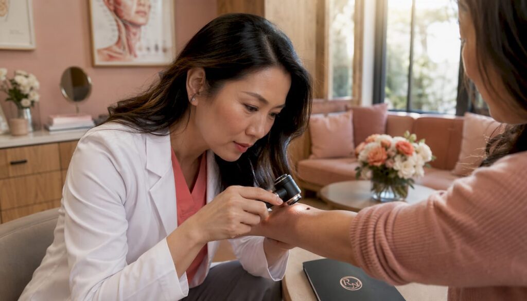
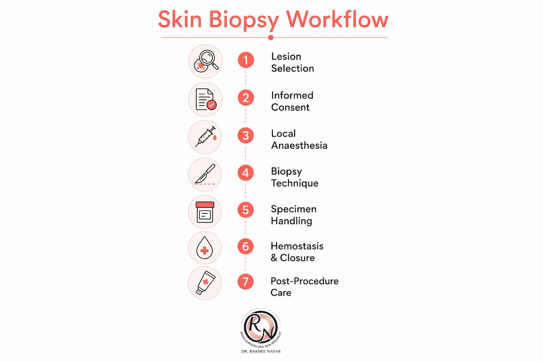
A skin biopsy obtains a tissue sample for histopathological analysis and, when performed systematically, gives the pathologist everything needed to reach a diagnosis. The core skin biopsy workflow runs as follows: select the correct lesion and site, obtain informed consent and complete safety checks, administer local anaesthesia, choose and execute the appropriate technique (punch, shave, saucerisation, incisional or excisional), handle and label the specimen correctly, achieve haemostasis and close the wound, then arrange documented follow-up once results are available. Guidance from NICE, the British Association of Dermatologists (BAD), and the British Society for Dermatological Surgery (BSDS) underpins each stage of this process.
Workflow at a glance:
- Lesion selection and clinical assessment
- Informed consent, allergies and anticoagulant checks, WHO/LocSSIP safety verification
- Local anaesthesia and patient preparation
- Technique selection and execution (punch, shave, incisional, excisional)
- Specimen handling, fixation and histopathology requisition
- Haemostasis and wound closure
- Post-procedure care and patient instructions
- Results communication and follow-up or referral
When should you biopsy, and which lesion should you choose?
The decision to biopsy should be driven by a clear clinical question. Common indications include diagnostic uncertainty about a pigmented or non-pigmented lesion, suspected malignancy (basal cell carcinoma, squamous cell carcinoma, melanoma), inflammatory dermatoses that have not responded to empirical treatment, and suspected immunobullous disease requiring direct immunofluorescence (DIF).
Site selection matters as much as the decision to biopsy. Sample the most representative, freshest lesion — not the oldest or most traumatised area. For blistering disorders, an intact early vesicle is preferred for light microscopy, while perilesional skin within 5–10 mm is recommended for DIF in most immunobullous conditions. For vasculitis, timing differs: early lesions (under 24 hours) are preferred for light microscopy, while DIF sampling may be taken from slightly older lesions. When a keratoacanthoma is suspected, sample the edge rather than the necrotic centre.
Relative contra-indications include active infection at the proposed site, uncorrected bleeding diathesis, and very fragile or atrophic skin where wound healing is likely to be poor. These are not absolute bars, but they alter technique and timing.
Critically, not every suspicious lesion warrants a biopsy in primary or intermediate care. Lesions meeting two-week wait referral criteria for suspected melanoma or SCC on the head and neck should generally be referred rather than sampled in a setting without the capacity for definitive management. A small incisional biopsy in primary care can delay care when excision is the appropriate first step.
Informed consent, documentation and the pre-procedure checklist
Consent for a skin biopsy must be specific, documented, and contemporaneous. Explain the indication, the alternatives (including watchful waiting or referral), and the risks: bleeding, infection, scarring, pigmentary change, and the possibility of a cosmetically suboptimal result. For facial or cosmetically sensitive sites, discuss the expected scar explicitly. Document all of this in the clinical record before the procedure begins.
Pre-procedure checklist:
- Confirm patient identity and the correct site (two-point verification)
- Check for known allergies, particularly to local anaesthetic agents and latex
- Review anticoagulant and antiplatelet therapy; apply BSDS antithrombotic guidance and CHA2DS2-VASc risk stratification where relevant
- Confirm pregnancy status where clinically relevant
- Inspect the site for local infection or skin breakdown
- Complete WHO surgical safety checklist and local LocSSIP requirements
- Photograph the lesion with orientation markers where feasible
The histopathology request form is a clinical document, not an administrative one. Include lesion duration, distribution, morphology, any recent topical or systemic treatment, your provisional differential diagnosis, and any specific questions for the pathologist (for example, whether DIF is required or whether ancillary microbiology is needed). A well-completed form meaningfully improves diagnostic yield, as StatPearls confirms.
Pro Tip: Use a standardised biopsy request template saved to your clinical system. Pre-populating the fixed fields (clinician name, GMC number, laboratory address) reduces omissions and saves time at the point of care.
Preparing patients before the procedure is covered in detail in the skin cancer biopsy preparation guide on the Rakhee Nayar – Mohs Surgeon and Skin Specialist website, which clinicians may find useful to share with patients in advance.
Local anaesthesia: agents, doses and infiltration technique
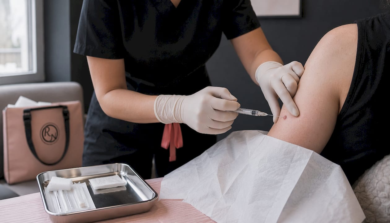
Lidocaine 1% or 2% with adrenaline 1:80,000 to 1:200,000 is the standard agent for most skin biopsies in adults. Adrenaline reduces bleeding and prolongs anaesthetic duration, but allow 15–20 minutes after infiltration before incising to achieve full vasoconstriction. Plain lidocaine without adrenaline is used where adrenaline is contra-indicated (end-arterial sites such as digits, or patient-specific factors), though the evidence base for absolute contra-indication at digital sites has evolved and local protocol should guide practice.
Infiltrate at the lesion margins using a field block rather than injecting directly into the lesion, which can distort architecture and compromise histological interpretation. Use a fine-gauge needle (27G or 30G) and aspirate before injecting in anatomically risky areas. Inject slowly to reduce discomfort; warming the solution to body temperature and buffering with sodium bicarbonate (typically 1:10 ratio) can further reduce the sting of infiltration.
Special scenarios require adjustment. Anticoagulated patients bleed more readily, so use the smallest effective volume and plan haemostasis in advance. Elderly patients with thin, atrophic skin need smaller volumes to avoid tissue distortion. For children, topical anaesthetic cream (EMLA or Ametop) applied under occlusion for 45–60 minutes before the procedure reduces procedural distress considerably.
Immediately before starting, confirm consent and site with the patient once more. Position the patient comfortably, ensure adequate lighting, and have an assistant available for larger or more complex biopsies.
Which biopsy technique should you use, and how do you perform it?
Technique selection follows the differential diagnosis. The table below maps common clinical scenarios to the appropriate method.
| Technique | Primary indications | Key sizing/depth guidance | Diagnostic limits |
|---|---|---|---|
| Punch biopsy | Inflammatory dermatoses, full-thickness sampling, suspected NMSC | 2–6 mm; bisect longitudinally if ≥4 mm | Limited for large or nodular lesions |
| Shave / saucerisation | Superficial or pedunculated lesions, seborrhoeic keratoses, some NMSC | Depth to mid-dermis at most | Understages melanoma; avoid if melanoma suspected |
| Incisional / wedge | Large lesions where excision is not feasible, suspected inflammatory disease | Include normal perilesional skin; orient to RSTL | Partial sample only |
| Excisional biopsy | Suspected melanoma, small lesions where complete removal is diagnostic and therapeutic | 1–2 mm margin; orient long axis to RSTL; full thickness to fat | Preferred for melanoma staging |
Punch biopsy
Stretch the skin perpendicular to relaxed skin tension lines (RSTLs) before punching; this produces an elliptical defect that closes more easily. Apply firm, rotating pressure with the punch, advancing to the subcutaneous fat. Lift the core gently with a needle or fine forceps, and cut the base with iris scissors rather than pulling, which causes crush artefact. Punches of 4 mm or larger should be bisected longitudinally to ensure representative sectioning; smaller cores can be embedded intact.
Shave and saucerisation
Use a flexible scalpel blade or a DermaBlade to shave at the appropriate depth. Saucerisation (a deeper, curved shave) samples into the reticular dermis and is suitable for suspected superficial BCC or actinic keratosis. Neither technique is appropriate when melanoma is in the differential, as transection of the base prevents accurate Breslow thickness measurement.
Excisional biopsy
For suspected melanoma, excisional biopsy with a 1–2 mm clinical margin is preferred where feasible, because Breslow thickness determines staging and subsequent management. Orient the long axis of the ellipse along RSTLs or lymphatic drainage lines where relevant. Carry the excision to the subcutaneous fat to avoid transecting the base. Mark orientation with a suture (for example, a short suture at the 12 o’clock pole) before placing the specimen in formalin.
Pro Tip: For biopsies on the eyelid, lip or nasal ala, plan the closure before you cut. These sites have limited tissue laxity and a poorly planned excision can produce ectropion, notching or distortion that is difficult to correct. If the lesion is large or the site is cosmetically critical, consider referral for facial reconstruction planning before proceeding.
Specimen handling, fixation and the histopathology requisition
Correct specimen handling begins the moment the tissue leaves the patient. Errors at this stage are irreversible.
Specimen handling checklist:
- Place routine specimens immediately into 10% neutral buffered formalin; do not allow drying
- For DIF, use Michel’s medium or transport in saline; never allow formalin contact with DIF specimens, as it destroys immunoglobulin deposits
- Use separate, clearly labelled containers for each specimen from different sites
- Mark orientation with a suture or ink before fixation; note the long axis and clock-face orientation on the request form
- Bisect punch cores ≥4 mm longitudinally; do not cut through intact vesicles, as the blister roof and contents carry diagnostic information
- Embed specimens with the skin surface perpendicular to the section plane
The histopathology request form should include: patient identifiers, site and laterality, lesion duration and morphology, clinical differential diagnosis, any recent treatment (topical steroids, imiquimod, cryotherapy), specific ancillary tests requested (DIF, microbiology, electron microscopy), and orientation details. Pathologists working with incomplete clinical information are more likely to issue a descriptive rather than a definitive report. The role of histopathology in skin cancer is explored in further detail on the Rakhee Nayar – Mohs Surgeon and Skin Specialist website for those who want a deeper understanding of what the pathologist does with the specimen.
A useful external resource on the clinical context that underpins good specimen labelling is this overview of common skin conditions, which reinforces why thorough clinical history improves diagnostic yield.
Haemostasis and wound closure: choosing the right approach
Most skin biopsies achieve haemostasis without difficulty. The method depends on wound size, site, and the patient’s anticoagulant status.
Haemostasis options:
- Direct pressure for 5–10 minutes: adequate for most small punch biopsies
- Chemical haemostasis: aluminium chloride 20% solution or Monsel’s solution (ferric subsulphate) for shave wounds and small punch sites; apply with a cotton-tipped applicator after drying the wound
- Electrocautery: effective for larger wounds but avoid on pedicled or random-pattern flaps; use sparingly near the eyes
- Topical tranexamic acid: an option for patients on anticoagulants where chemical agents are insufficient, though local protocol should guide use
Closure choices follow wound size and site. Small punch biopsies (3–4 mm) on the trunk or limbs often close satisfactorily with a single interrupted suture or adhesive strip. Facial punch sites generally warrant a 5/0 or 6/0 non-absorbable monofilament suture (nylon or polypropylene) to minimise scarring. Larger excisional wounds benefit from a layered closure: absorbable sutures (for example, 3/0 or 4/0 Vicryl Rapide) for the deep layer, and non-absorbable monofilament for the epidermis.
Per BSDS 2023 antithrombotic guidance, most small skin biopsies can proceed without stopping direct oral anticoagulants (DOACs). Once haemostasis is secured, DOACs can usually be restarted within 24 hours. For patients at higher thromboembolic risk, consult the prescriber or haematology before any interruption. Patients on anticoagulants warrant observation after the procedure where there is clinical concern about bleeding.
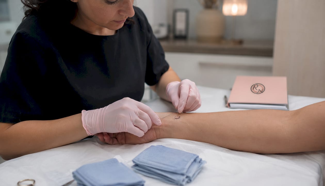
Apply a non-adherent primary dressing and a compression dressing where appropriate. Advise elevation of the limb for 24–48 hours if the biopsy is on the lower leg.
Post-procedure care and complications to counsel patients about
Standard aftercare is straightforward. Keep the dressing dry for 48 hours, avoid strenuous activity during that period, and elevate the site where possible. After 48 hours, patients may clean the wound gently with saline or clean water and reapply a simple dressing until the wound is healed or sutures are removed.
Suture removal timing varies by site: facial sutures at 5–7 days, scalp at 7–10 days, trunk and upper limb at 10–14 days, and lower limb at 14 days or longer, particularly in elderly patients or those with poor peripheral circulation.
Complications are uncommon. Infection rates are below 1% for routine skin biopsies, and prophylactic antibiotics are not indicated as standard practice. Counsel patients about the signs of wound infection (increasing redness, warmth, purulent discharge, fever) and advise them to seek review promptly if these develop. Haematoma, wound dehiscence, and hypertrophic scarring are the other complications to mention at consent. Sensory changes around the biopsy site are common and usually temporary.
Prophylactic antibiotics may be appropriate for immunocompromised patients, those with prosthetic heart valves (following current NICE guidance), or where the wound is in a high-risk anatomical area. Follow local formulary guidance.
How do you manage results and arrange appropriate follow-up?
Laboratory turnaround time for histopathology varies by local trust or private laboratory. Counsel patients that results are not immediate and that they will be contacted once the report has been reviewed clinically. A reasonable expectation in most NHS settings is 2–4 weeks; private laboratories often report faster.
A simple follow-up workflow by result:
- Benign result, diagnosis confirmed: inform the patient, discharge or continue routine surveillance as appropriate, and document the result in the record.
- Premalignant lesion (actinic keratosis, Bowen’s disease, lentigo maligna): discuss treatment options, arrange appropriate therapy or referral, and document the management plan.
- Malignancy requiring further excision (BCC, SCC, invasive melanoma): arrange definitive treatment promptly. For BCC or SCC on the face or cosmetically sensitive sites, consider referral for Mohs micrographic surgery where margin control is a priority.
- Melanoma: urgent referral to a skin cancer multidisciplinary team (MDT). Breslow thickness, ulceration, and mitotic rate from the report determine the next steps. Do not delay while awaiting a clinic appointment; contact the MDT directly.
- Inconclusive or non-diagnostic report: review the clinical context, consider whether the biopsy site was representative, and discuss with the reporting pathologist. A repeat biopsy or specialist referral may be needed.
Document all results communication in the clinical record, including the date, the method of communication, and the agreed management plan.
When should you refer rather than biopsy?
Some lesions should not be biopsied in a non-specialist setting. Knowing when to stop and refer is as important as knowing how to perform the procedure.
Red flags for urgent two-week wait referral:
- Any lesion with features of melanoma (asymmetry, border irregularity, colour variation, diameter >6 mm, evolving morphology) where excision rather than incisional sampling is the appropriate first step
- Indurated or ulcerated lesions >1 cm on the head or neck suspicious for SCC
- Rapidly changing lesions, particularly in immunosuppressed patients
- Lesions where the clinical presentation strongly suggests a diagnosis that requires definitive excision rather than diagnostic sampling
Situations where biopsy may delay care:
A small incisional biopsy of a large lesion suspicious for melanoma can delay definitive management and, if the sample is not representative, produce a falsely reassuring result. Where urgent referral is clinically indicated, refer via the two-week wait pathway rather than attempting in-house sampling.
When to seek specialist advice before proceeding:
- Challenging anatomical sites (eyelid margin, lip vermilion, nasal tip, ear helix) where a poorly planned biopsy can cause functional or cosmetic harm
- Patients with significant comorbidity, complex anticoagulation, or immunosuppression
- Suspected immunobullous disease where DIF expertise and appropriate transport media are needed
- Any case where the clinician is uncertain about technique, orientation, or specimen handling
Referral checklist:
- Document the clinical indication and urgency clearly in the referral
- Include lesion photographs with a scale marker where possible
- State whether a biopsy has already been attempted and, if so, include the histopathology report
- Specify the two-week wait pathway where criteria are met
- Communicate directly with the receiving team for high-risk or complex cases
Consultant-level tips to reduce diagnostic error and improve cosmetic results
A few habits separate a technically adequate biopsy from one that gives the pathologist exactly what they need.
Photograph every lesion before infiltrating local anaesthetic. Anaesthetic infiltration blanches the skin and can obscure colour variation that was diagnostically relevant. A photograph taken in good lighting, with a ruler in frame and an orientation marker, becomes part of the clinical record and helps the pathologist correlate the macroscopic appearance with the histology.
When marking orientation, use a consistent system and record it on both the specimen container and the request form. A short suture at 12 o’clock is reliable and survives formalin fixation. Ink marks applied before excision can be useful but may wash off during processing if not fixed.
For paired biopsies (one in formalin, one for DIF), label the containers in the room before either specimen is placed. Formalin contamination of a DIF specimen is an irreversible error. Consider a verbal read-back with your assistant: “This container is Michel’s medium, this one is formalin.”
Minimising diagnostic error also means annotating the request form with features the pathologist cannot see: the dermoscopic pattern, the rate of change, the patient’s immunosuppression status, or a recent course of topical steroids that may have modified the histology.
For cosmetic optimisation, the single most effective adjustment is correct orientation. An ellipse placed along RSTLs heals with a finer scar than one placed across them. On the face, plan the long axis before infiltrating, not after.
Pro Tip: Before making the first incision, pause and perform a verbal WHO check with your assistant: patient name, site, procedure, and any known allergies. Wrong-site biopsy is a recognised Never Event in dermatology. This 30-second habit, mandated by BSDS practice standards, costs nothing and prevents an irreversible error.
Key takeaways
A safe, diagnostically useful skin biopsy requires correct lesion selection, complete pre-procedure documentation, appropriate technique, and meticulous specimen handling — failure in any one of these domains reduces the value of the procedure.
| Point | Details |
|---|---|
| Select the right lesion and site | Sample the freshest, most representative lesion; use perilesional skin within 5–10 mm for DIF. |
| Complete safety checks every time | WHO checklist and LocSSIP adherence prevent wrong-site biopsy, a recognised Never Event in dermatology. |
| Match technique to differential | Excisional biopsy is preferred for suspected melanoma; shave technique understages and should be avoided. |
| Specimen handling is irreversible | DIF specimens must never contact formalin; punch cores ≥4 mm should be bisected longitudinally. |
| Rakhee Nayar – Mohs Surgeon and Skin Specialist | Specialist assessment at Circle Cheshire for complex sites, suspected melanoma, or where reconstructive planning is needed after biopsy. |
Why specimen quality matters more than technique speed
There is a tendency in busy clinical practice to treat the biopsy as the end of the diagnostic process. It is not. The biopsy is the beginning of a conversation with the pathologist, and the quality of that conversation depends almost entirely on what happens before the specimen reaches the laboratory.
The clinicians who get the most useful reports are not necessarily the most technically gifted. They are the ones who complete the request form properly, photograph the lesion before anaesthesia, mark orientation consistently, and choose the right transport medium without being prompted. These habits take thirty seconds each. Collectively, they are the difference between a report that says “consistent with” and one that gives a definitive diagnosis with clear margins.
The other thing worth saying plainly: knowing when not to biopsy is a clinical skill. Referring a lesion that meets two-week wait criteria, rather than sampling it in a setting without the capacity for definitive management, is the right decision. A biopsy that delays a melanoma diagnosis by three weeks is not a neutral act.
When to consider a specialist consultation at Circle Cheshire
For lesions on cosmetically sensitive sites, suspected melanoma, or cases where the biopsy result points towards a treatment that requires specialist input, a consultation with Miss Rakhee Nayar at Circle Cheshire offers a clear next step. Miss Nayar is a GMC-registered Consultant Plastic Surgeon, FRCS (Plast), MD, dual-trained in plastic surgery and Mohs micrographic surgery, and sees patients for both diagnostic assessment and definitive treatment.

Private consultation fees and biopsy costs at Circle Cheshire are available on request; typical private initial consultation fees in the North West range from £200–£350, with procedural costs discussed at the time of booking. For lesions where Mohs surgery may be the most appropriate treatment, early specialist involvement avoids the need for staged procedures later. For patients or referring clinicians who want to understand the full pathway from biopsy to skin cancer detection and treatment, the clinic offers both in-person and e-consultation appointments.
To arrange a referral or consultation, contact the clinic directly via mohssurgeon.co.uk. Please note: this article is general clinical information and does not constitute medical advice. Clinicians and patients should confirm management decisions with a GMC-registered specialist for their individual circumstances.
Useful sources and recommended reading
| Source | Practical utility |
|---|---|
| StatPearls: Skin Biopsy | Stepwise technique overview, haemostasis, infection rates, and clinical history guidance |
| RCPath: Tissue pathways for dermatopathology | Specimen handling, fixation media, DIF transport, bisection rules, embedding orientation |
| BSDS: Antithrombotics and skin surgery | Anticoagulant management, DOAC continuation, CHA2DS2-VASc stratification, turnaround counselling |
| BAD: Serious incidents and Never Events | Wrong-site surgery prevention, LocSSIP and WHO checklist requirements |
| BSDS: Online learning and practice standards | Safety culture, checklist adherence, procedural standards for dermatological surgery |
| NEJM: How to perform a punch biopsy | Video and stepwise guide to punch biopsy technique |
| PMC: Techniques of skin biopsy | Comparative technique review including shave, punch, incisional and excisional methods |
Clinicians should also follow their local LocSSIPs and the NHS England WHO Surgical Safety Checklist for all invasive dermatology procedures, however minor.
FAQ
What is the step-by-step process of a skin biopsy?
The skin biopsy procedure follows seven steps: lesion selection and clinical assessment, informed consent and safety checks, local anaesthesia, technique selection and execution (punch, shave, incisional or excisional), specimen handling and fixation, haemostasis and wound closure, and documented follow-up once histopathology results are available.
How long does skin biopsy histopathology take to process?
Turnaround time varies by laboratory. NHS trusts typically report within 2–4 weeks; private laboratories often report faster. Patients should be counselled that results follow clinical review and that timelines are not fixed.
How painful is a skin biopsy?
The procedure itself is performed under local anaesthesia and should cause minimal discomfort once the anaesthetic has taken effect. The injection of lidocaine can sting briefly; buffering the solution and injecting slowly reduces this. Some soreness is expected for 24–48 hours afterwards.
What should patients avoid after a skin biopsy?
Patients should keep the dressing dry for 48 hours, avoid strenuous activity during that period, and refrain from submerging the wound (swimming, baths) until it is fully healed. They should seek review if they notice signs of infection, increasing pain, or bleeding that does not settle with direct pressure.
When is referral preferable to an in-clinic biopsy?
Lesions meeting two-week wait criteria for suspected melanoma or SCC on the head and neck, large or rapidly changing lesions, and cases on cosmetically sensitive anatomical sites should generally be referred rather than sampled in a non-specialist setting. Miss Nayar at Circle Cheshire accepts referrals for complex or high-risk cases via mohssurgeon.co.uk.
Recommended
- Skin cancer symptoms and signs: the UK guide for early detection
- How to prepare for a skin cancer biopsy
- Skin cancer specialist referral criteria: a UK patient guide
- Role of histopathology in skin cancer: a clinical guide
Filed under Spotting Skin Cancer


